. 2003 Apr 1;100(7):4114-9.
doi: 10.1073/pnas.0633863100. Epub 2003 Mar 17.

Bifunctional antisense oligonucleotides provide a trans-acting splicing enhancer that stimulates SMN2 gene expression in patient fibroblasts

Affiliations

Bifunctional antisense oligonucleotides provide a trans-acting splicing enhancer that stimulates SMN2 gene expression in patient fibroblasts

Leigh A Skordis et al. Proc Natl Acad Sci U S A. .

Abstract

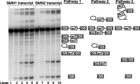
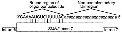
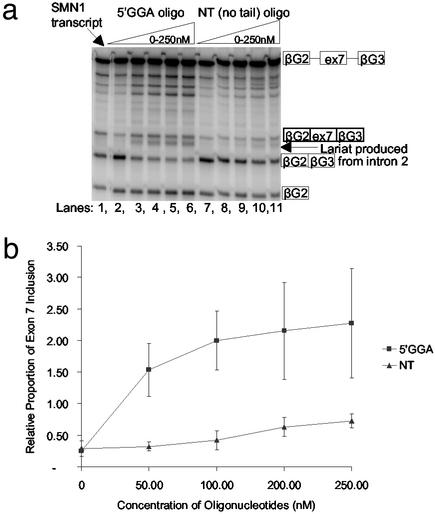
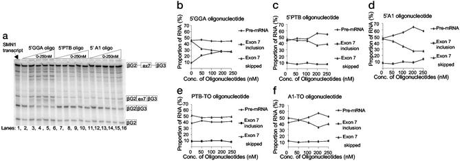
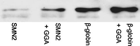
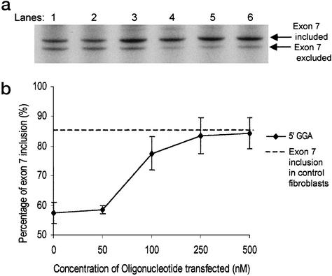
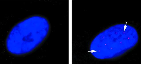
The multiplicity of proteins compared with genes in mammals owes much to alternative splicing. Splicing signals are so subtle and complex that small perturbations may allow the production of new mRNA variants. However, the flexibility of splicing can also be a liability, and several genetic diseases result from single-base changes that cause exons to be skipped during splicing. Conventional oligonucleotide strategies can block reactions but cannot restore splicing. We describe here a method by which the use of a defective exon was restored. Spinal muscular atrophy (SMA) results from mutations of the Survival Motor Neuron (SMN) gene. Mutations of SMN1 cause SMA, whereas SMN2 acts as a modifying gene. The two genes undergo alternative splicing with SMN1, producing an abundance of full-length mRNA transcripts, whereas SMN2 predominantly produces exon 7-deleted transcripts. This discrepancy is because of a single nucleotide difference in SMN2 exon 7, which disrupts an exonic splicing enhancer containing an SF2ASF binding site. We have designed oligoribonucleotides that are complementary to exon 7 and contain exonic splicing enhancer motifs to provide trans-acting enhancers. These tailed oligoribonucleotides increased SMN2 exon 7 splicing in vitro and rescued the incorporation of SMN2 exon 7 in SMA patient fibroblasts. This treatment also resulted in the partial restoration of gems, intranuclear structures containing SMN protein that are severely reduced in patients with SMA. The use of tailed antisense oligonucleotides to recruit positively acting factors to stimulate a splicing reaction may have therapeutic applications for genetic disorders, such as SMA, in which splicing patterns are altered.


Figures

Figure 1
Figure 1
In vitro splicing assay incorporating SMN1 and SMN2 transcripts shows alternative splicing. Denaturing polyacrylamide gel (5%) showing an in vitro timed splicing assay using α-32P-labeled SMN1 and SMN2 transcripts. Symbols identifying the various splicing intermediates and products are shown. Lanes 1–5 and 6–10 represent different time points. Lanes 1 and 6, 0 min; lanes 2 and 7, 30 min; lanes 3 and 8, 1 h; lanes 4 and 9, 2 h; lanes 5 and 10, 3 h. Also shown are the three different splicing pathways that occur; pathways 1 and 2 promote exon 7 inclusion, whereas the third pathway skips exon 7.
Figure 2
Figure 2
Diagrammatical representation of the tailed oligoribonucleotides bound to SMN2 exon 7. The complementary RNA sequence of the oligoribonucleotide is uppercase, whereas the tail region containing sequences that mimic ESEs are lowercase. The oligonucleotide binds via a complementary region to the first part of SMN2 exon 7 (diagram not to scale) indicated by the vertical lines, from position +1 on exon 7 to position +16, thereby leaving 38 nucleotides of the exon unannealed by the oligonucleotide. The noncomplementary tail region remains unbound and is available to bind to splicing proteins in the in vitro splicing reaction mix.
Figure 3
Figure 3
Tailed 5′GGA oligonucleotides promote exon 7 inclusion. (a) Cell-free in vitro splicing assay using α-32P-labeled SMN2 transcripts combined with the 5′GGA and NT oligonucleotides at concentrations between 0 and 250 nM. The SMN1 transcript is included in the first lane. (b) Graph showing exon 7 inclusion relative to the SMN1 level of splicing with increasing concentrations of oligonucleotides. The SMN1 transcript was included in all gels as an internal control, enabling successive gels to be directly correlated. The “relative proportion” label on the y axis indicates that the readings have been normalized against the readings obtained for the SMN1 transcripts in each gel. The results of three experiments were combined to produce these data with SDs varying from 0.03 to 0.86. The readings were not corrected for the differing amounts of labeled radionucleotides.
Figure 4
Figure 4
Application of the 5′GGA, 5′PTB, 5′A1, PTB-TO, and A1-TO oligonucleotides to SMN2 transcripts. (a) Denaturing polyacrylamide gel (5%) with 5′GGA, 5′PTB, and 5′A1 oligonucleotides incorporated at increasing concentrations with the SMN2 transcripts. The SMN1 transcript is included in the first lane. (bf) Graphs showing the percentage of RNA in the initial pre-mRNA transcript, the exon 7-included product, and the skipped product at increasing concentrations of each separate oligonucleotide. The products have been corrected for the numbers of labeled radionucleotides in each form of RNA. These graphs were plotted from a single experiment, but the results were reproducible in at least three different experiments; however, the error bars have been excluded to avoid confusion of the results shown.
Figure 5
Figure 5
Recruitment of SF2/ASF to SMN2 exon 7 by the 5′GGA oligonucleotide. Biotinylated RNA (SMN2 exon 7 or β-globin, as indicated) was bound to streptavidin beads and incubated in nuclear extract. The proteins associated with the RNA were separated by SDS/PAGE, and SF2/ASF was detected by Western blotting. Lanes “+GGA” indicate that the RNA was incubated with the GGA oligonucleotide before addition to the beads.
Figure 6
Figure 6
Transfection of type I SMA patient fibroblasts. (a) Denaturing polyacrylamide gel (6%) showing the results of a semiquantitative RT-PCR, using primers situated in exons 6 and 8 of the SMN gene, was carried out on cDNA from cells transfected with increasing concentrations of the 5′GGA oligonucleotide. Lane 1, untransfected cells; lane 2, 50 nM oligonucleotide-transfected; lane 3, 100 nM oligonucleotide-transfected; lane 4, 250 nM oligonucleotide-transfected; lane 5, 500 nM oligonucleotide-transfected; lane 6, normal control. (b) Graph showing the percentage of exon 7 inclusion in the transcripts derived from the 5′GGA transfected cells. The results of three different transfection experiments were combined to produce the graph. The readings were corrected for the amounts of labeled radionucleotides, and the percentage of exon 7 inclusion was calculated by exon 7 inclusion mRNA/total mRNA. The horizontal dashed line represents the percentage of exon 7 inclusion obtained in control fibroblasts.
Figure 7
Figure 7
Restoration of gems in SMA patient fibroblasts. Images show untransfected (a) and transfected (b) SMA type I fibroblasts. 4′,6-Diamidino-2-phenylindole staining highlights the nuclei in blue, and the white arrows indicate the gems (red dots in nucleus). Untransfected cells show 2–3% of their nuclei containing gems, whereas transfected cells show 13% gem-positive nuclei.

Comment in

References

    1. Dubowitz V. Muscle Disorders in Childhood. 2nd Ed. London: Saunders; 1995.
    1. Lefebvre S, Burglen L, Reboullet S, Clermont O, Burlet P, Viollet L, Benichou B, Cruaud C, Millasseau P, Zeviani M. Cell. 1995;80:155–165. - PubMed
    1. Liu Q, Dreyfuss G. EMBO J. 1996;15:3555–3565. - PMC - PubMed
    1. McAndrew P E, Parsons D W, Simard L R, Rochette C, Ray P N, Mendell J R, Prior T W, Burghes A H M. Am J Hum Genet. 1997;60:1411–1422. - PMC - PubMed
    1. Taylor J E, Thomas N H, Lewis C M, Abbs S J, Rodrigues N R, Davies K E, Mathew C G. Eur J Hum Genet. 1998;6:467–474. - PubMed

Publication types

MeSH terms