. 2003 Apr;185(7):2096-103.
doi: 10.1128/JB.185.7.2096-2103.2003.

OmcB, a c-type polyheme cytochrome, involved in Fe(III) reduction in Geobacter sulfurreducens

Affiliations

OmcB, a c-type polyheme cytochrome, involved in Fe(III) reduction in Geobacter sulfurreducens

Ching Leang et al. J Bacteriol. 2003 Apr.

Abstract

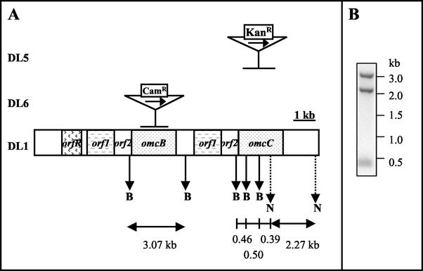
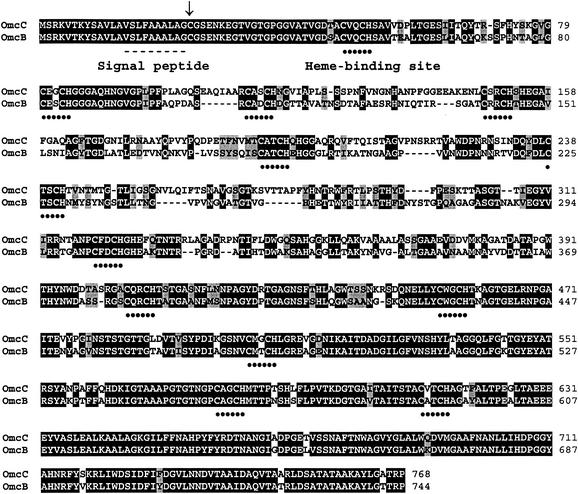
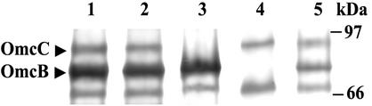
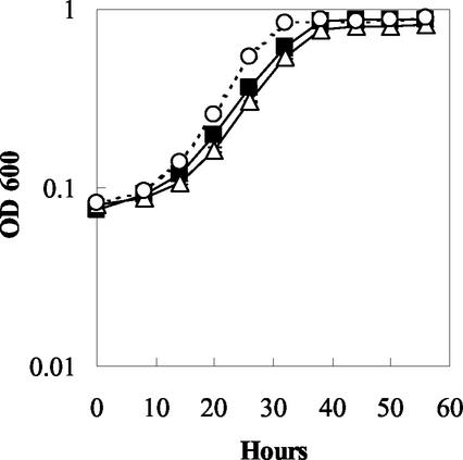
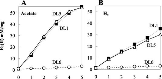
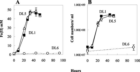
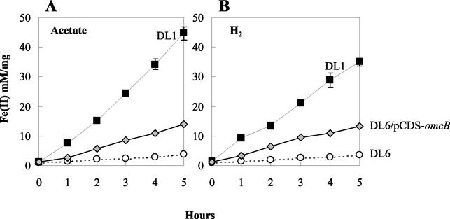
Microorganisms in the family Geobacteraceae are the predominant Fe(III)-reducing microorganisms in a variety of subsurface environments in which Fe(III) reduction is an important process, but little is known about the mechanisms for electron transport to Fe(III) in these organisms. The Geobacter sulfurreducens genome was found to contain a 10-kb chromosomal duplication consisting of two tandem three-gene clusters. The last genes of the two clusters, designated omcB and omcC, encode putative outer membrane polyheme c-type cytochromes which are 79% identical. The role of the omcB and omcC genes in Fe(III) reduction in G. sulfurreducens was investigated. OmcB and OmcC were both expressed during growth with acetate as the electron donor and either fumarate or Fe(III) as the electron acceptor. OmcB was ca. twofold more abundant under both conditions. Disrupting omcB or omcC by gene replacement had no impact on growth with fumarate. However, the OmcB-deficient mutant was greatly impaired in its ability to reduce Fe(III) both in cell suspensions and under growth conditions. In contrast, the ability of the OmcC-deficient mutant to reduce Fe(III) was similar to that of the wild type. When omcB was reintroduced into the OmcB-deficient mutant, the capacity for Fe(III) reduction was restored in proportion to the level of OmcB production. These results indicate that OmcB, but not OmcC, has a major role in electron transport to Fe(III) and suggest that electron transport to the outer membrane is an important feature in Fe(III) reduction in this organism.


Figures

FIG. 1.
FIG. 1.
Organization of the gene duplication in G. sulfurreducens and mutation schemes. (A) Two gene clusters, orf1/orf2/omcB and orf1/orf2/omcC, were identified within a 10-kb duplication region in G. sulfurreducens (DL1). The central part of either omcC or omcB was replaced with a kanamycin or a chloramphenicol cassette in the mutant DL5 (omcC::kan) or DL6 (omcB::cam), respectively. Gene replacement is indicated by a horizontal bar. The transcription direction of the Kanr and Camr cassettes is the same of that of the omcC and omcB genes and is indicated by a horizontal arrow. A partial restriction enzyme map is shown with vertical arrows indicating BamHI sites (capital B) and NcoI sites (capital N; dotted line). (B) Southern blot of genomic DNA prepared from wild-type G. sulfurreducens (DL1). Genomic DNA digested with restriction enzymes BamHI and NcoI was probed with the omcC gene amplified with primers 8901 and 8904 (primer sequences listed in Materials and Methods). Expected radiolabeled bands are 2.27, 0.5, 0.46, and 0.39 kb. However, due to the high identity between omcB and omcC, an extra band of 3.07 kb, corresponding to the omcB gene, was also detected in the blot. The blot analysis proves that both omcB and omcC genes exist in the G. sulfurreducens genome. Restriction map was based on sequence data obtained from The Institute for Genetic Research website (http://www.tigr.org).
FIG. 2.
FIG. 2.
Amino acid sequence alignments of OmcB and OmcC. Identical and conservatively substituted residues are highlighted in black and gray, respectively. Heme-binding domains (CXXCH) are indicated with dotted lines. The signal peptide (dashed line) and lipid attachment site (arrow) are indicated for both proteins.
FIG. 3.
FIG. 3.
Heme-staining and Tricine-polyacrylamide gel electrophoresis of membrane fractions prepared from DL1, DL5 (omcC::kan), DL6 (omcB::cam), and the complemented strain DL6/pCDS-omcB. Membrane fractions were prepared from cultures grown with acetate-fumarate (lane 1, DL1; lane 3, DL5; lane 4, DL6) or acetate-Fe(III) citrate (lane 2, DL1; lane 5, DL6/pCDS-omcB). Membrane proteins (50 μg) were resolved on a 10% Tris-Tricine polyacrylamide gel and stained for heme. OmcB and OmcC are indicated with arrows.
FIG. 4.
FIG. 4.
Growth study showing results for DL1 (filled square), DL5 (omcC::kan) (empty triangle), and DL6 (omcB::cam) (empty circle) in the medium containing acetate as the electron donor and fumarate as the electron acceptor. Cell growth was measured at A600 over time. Data are means ± standard deviations (SD) of triplicates.
FIG. 5.
FIG. 5.
Cell suspension assay showing the production of Fe(II) by DL1 and mutants DL5 (omcC::kan) and DL6 (omcB::cam). Each sample was assayed in triplicate, and all assays were done in parallel. Acetate (A) or hydrogen (B) was supplied as the electron donor. Symbols: DL1 (filled square); DL5 (empty triangle); DL6 (empty circle). Data are means ± SD of triplicates.
FIG. 6.
FIG. 6.
Growth of DL1 (filled square), DL5 (empty triangle), and DL6 (empty circle) in medium containing acetate as the electron donor and Fe(III) citrate as the electron acceptor. Mid-log (A600 = ∼0.3) fumarate-grown cells were inoculated (3% inoculum) into fresh Fe(III) citrate medium. (A) Fe(II) production; (B) cell density. Data are means ± SD of triplicates.
FIG. 7.
FIG. 7.
Cell suspensions assay showing the production of Fe(II) by DL1 and mutants DL6 and the complemented strain DL6/pCDS-omcB. Each sample was assayed in triplicate, and all assays were done in parallel. Acetate (A) or hydrogen (B) was supplied as the electron donor. Symbols: DL1 (filled square); DL6 (empty circle); DL6/pCDS-omcB (triangle shaded in gray). Data are means ± SD of triplicates.

References

    1. Ausubel, F. M., R. Brent, R. E. Kingston, D. D. Moore, J. G. Seidman, J. A. Smith, and K. Struhl (ed.). 1999. Current protocols in molecular biology, vol. 2. John Wiley & Sons, Inc., New York, N.Y.
    1. Beliaev, A., D. Saffarini, J. McLaughlin, and D. Hunnicutt. 2001. MtrC, an outer membrane decahaem c cytochrome required for metal reduction in Shewanella putrefaciens MR-1. Mol. Microbiol. 39:722-730. - PubMed
    1. Beliaev, A. S., and D. A. Saffarini. 1998. Shewanella putrefaciens mtrB encodes an outer membrane protein required for Fe(III) and Mn(IV) reduction. J. Bacteriol. 180:6292-6297. - PMC - PubMed
    1. Caccavo, F., Jr., D. J. Lonergan, D. R. Lovley, M. Davis, J. F. Stolz, and M. J. McInerney. 1994. Geobacter sulfurreducens sp. nov., a hydrogen- and acetate-oxidizing dissimilatory metal-reducing microorganism. Appl. Environ. Microbiol. 60:3752-3759. - PMC - PubMed
    1. Chang, A. C., and S. N. Cohen. 1978. Construction and characterization of amplifiable multicopy DNA cloning vehicles derived from the P15A cryptic miniplasmid. J. Bacteriol. 134:1141-1156. - PMC - PubMed

Publication types

MeSH terms