. 2003 Apr;185(7):2296-305.
doi: 10.1128/JB.185.7.2296-2305.2003.

Evidence for horizontal transfer of the EcoT38I restriction-modification gene to chromosomal DNA by the P2 phage and diversity of defective P2 prophages in Escherichia coli TH38 strains

Affiliations

Evidence for horizontal transfer of the EcoT38I restriction-modification gene to chromosomal DNA by the P2 phage and diversity of defective P2 prophages in Escherichia coli TH38 strains

Keiko Kita et al. J Bacteriol. 2003 Apr.

Abstract

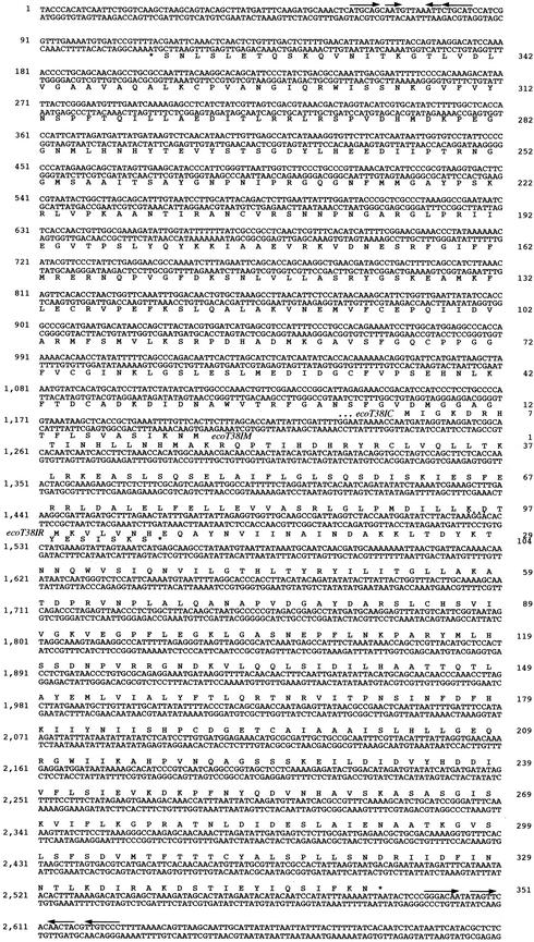
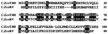
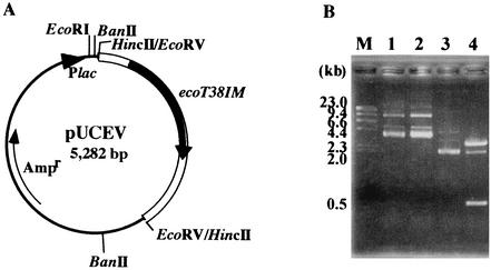
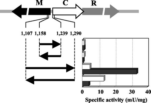
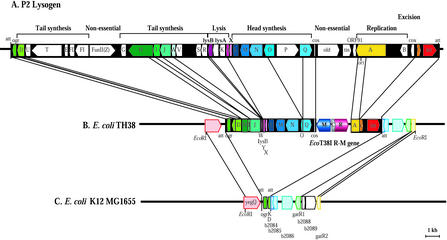
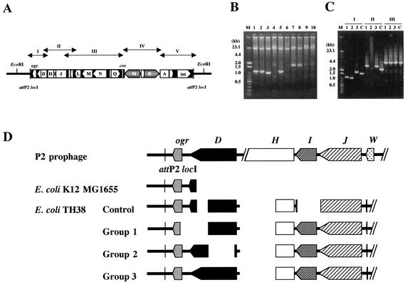
A DNA fragment carrying the genes coding for a novel EcoT38I restriction endonuclease (R.EcoT38I) and EcoT38I methyltransferase (M.EcoT38I), which recognize G(A/G)GC(C/T)C, was cloned from the chromosomal DNA of Escherichia coli TH38. The endonuclease and methyltransferase genes were in a head-to-head orientation and were separated by a 330-nucleotide intergenic region. A third gene, the C.EcoT38I gene, was found in the intergenic region, partially overlapping the R.EcoT38I gene. The gene product, C.EcoT38I, acted as both a positive regulator of R.EcoT38I gene expression and a negative regulator of M.EcoT38I gene expression. M.EcoT38I purified from recombinant E. coli cells was shown to be a monomeric protein and to methylate the inner cytosines in the recognition sequence. R.EcoT38I was purified from E. coli HB101 expressing M.EcoT38I and formed a homodimer. The EcoT38I restriction (R)-modification (M) system (R-M system) was found to be inserted between the A and Q genes of defective bacteriophage P2, which was lysogenized in the chromosome at locI, one of the P2 phage attachment sites observed in both E. coli K-12 MG1655 and TH38 chromosomal DNAs. Ten strains of E. coli TH38 were examined for the presence of the EcoT38I R-M gene on the P2 prophage. Conventional PCR analysis and assaying of R activity demonstrated that all strains carried a single copy of the EcoT38I R-M gene and expressed R activity but that diversity of excision in the ogr, D, H, I, and J genes in the defective P2 prophage had arisen.


Figures

FIG.1.
FIG.1.
Nucleotide sequence of the 2,700-bp region. The amino acid sequences assigned to the C.EcoT38I (ecoT38IC) and R.EcoT38I (ecoT38IR) genes are given below the nucleotide sequences, and that assigned to the M.EcoT38I (ecoT38IM) gene is given above the nucleotide sequence. The nucleotide sequence is numbered from the leftmost end, and the deduced amino acid sequences of R.EcoT38I, M.EcoT38I, and C.EcoT38I are numbered from the initiation codon of each gene. The potential ribosome-binding sequence is indicated by dots. Pairs of arrows indicate palindromic sequences characteristic of the termination signal.
FIG. 2.
FIG. 2.
Alignment of C.EcoT38I and C.EcoRV. Each line represents one sequence. Shading indicates conserved amino acid residues. Gaps in the aligned sequences are indicated by dashes.
FIG. 3.
FIG. 3.
Analysis of the methylation specificity of M.EcoT38I (ecoT38IM). (A) Structure of pUCEV, in which the 2.2-kb EcoRV fragment carrying the M.EcoT38I gene was cloned into the HincII site of pUC118. (B) Resistance to R.BanII of M.EcoT38I-modified DNA. pUCEV and pUC118 were incubated with R.BanII. The products were separated by 1% agarose gel electrophoresis. Lane 1, pUCEV; lane 2, pUCEV plus R.BanII; lane 3, pUC118; lane 4, pUC118 plus R.BanII. λ HindΙΙΙ digests were used for molecular size calibration (lane M).
FIG. 4.
FIG. 4.
Effect of C.EcoT38I on the promoter activities of the R.EcoT38I and M.EcoT38I genes. The segments from 1,158 to 1,239 bp and from 1,107 to 1,290 bp shown in Fig. 1 were cloned upstream of a promoterless AR1 gene in vector pMCLTerAR. Promoter activities associated with various DNA fragments were measured for both orientations. Each clone was assayed for AR1 activity in the absence (open bars) and presence (closed bars) of C.EcoT38I (graph). R, M, and C represent R.EcoT38I, M.EcoT38I, and C.EcoT38I, respectively.
FIG. 5.
FIG. 5.
Gene organization of the EcoT38I R-M system and flanking regions on the E. coli TH38 chromosome. (A) Gene order of the P2 phage integrated into the E. coli chromosome. (B) Gene order of the 16-kb EcoRI region flanking the EcoT38I R-M genes on the E. coli TH38 chromosome. The genes and their directions of transcription, cos, and phage attachment sites (att) are shown. R, M, and C represent R.EcoT38I, M.EcoT38I, and C.EcoT38I, respectively. (C) Gene order of P2 att (locI) and flanking regions in E. coli K-12 MG1655. att indicates att (locI).
FIG. 6.
FIG. 6.
Variability of defective P2 prophage genomes in E. coli TH38 DNA. (A) Locations of target DNAs for PCR amplification. (B) Agarose gel electrophoresis of PCR products obtained from E. coli TH38 strains with primers I61 and I62. The PCR mixture (50 μl) comprised 25 pmol of each primer, 400 μM each deoxynucleotide triphosphate, 2.5 mM MgCl2, LA-PCR buffer (Mg2+ free), template DNA, and 2.5 U of LA Taq DNA polymerase. The reaction mixture was overlaid with mineral oil, and the reaction was carried out with a Perkin-Elmer Cetus thermal cycler. The initial template denaturation step consisted of 1 min at 94°C. The amplification profile (20 s at 98°C and 25 min at 68°C) was repeated for 30 cycles. Lanes 1 to 10 correspond to products from 10 E. coli TH38 colonies picked randomly. A mixture of λ HindIII digests and 100-bp DNA ladder markers (Toyobo) was used for molecular size calibration (lane M). The PCR products were subjected to 1% agarose gel electrophoresis and then stained with ethidium bromide. (C) Agarose gel electrophoresis of E. coli DNA fragments amplified by PCR with primers A and B (I), C and D (II), and E and F (III). The PCR mixture (50 μl) comprised 10 pmol of each primer, 200 μM each deoxynucleotide triphosphate, 2.5 mM MgCl2, LA-PCR buffer (Mg2+ free), template DNA, and 2.5 U of LA Taq DNA polymerase. The reaction mixture was overlaid with mineral oil, and the reaction was carried out with a Perkin-Elmer Cetus thermal cycler. The initial template denaturation step consisted of 1 min at 94°C. The amplification profile (30 s at 94°C, 1 min at 60°C, and 5 min at 72°C) was repeated for 25 cycles. Lanes C correspond to the products from the E. coli TH38 control strain, for which the complete nucleotide sequence of the 16-kb EcoRI region was determined in this study. Lanes 1 to 3 correspond to products with different molecular sizes from the E. coli TH38 strains analyzed in panel B. (D) Schematic diagram of the defective P2 prophage genome in various E. coli TH38 strains. Groups 1 to 3 correspond to lanes 1 to 3 in panel C.

Similar articles

Cited by

References

    1. Anton, B. P., D. F. Heiter, J. S. Benner, E. J. Hess, L. Greenough, L. S. Moran, B. E. Slatko, and J. E. Brooks. 1997. Cloning and characterization of the BglII restriction-modification system reveals a possible evolutionary footprint. Gene 187:19-27. - PubMed
    1. Barreiro, V., and E. Haggard-Ljungquist. 1992. Attachment sites for bacteriophage P2 on the Escherichia coli chromosome: DNA sequences, localization on the physical map, and detection of a P2-like remnant in E. coli K-12 derivatives. J. Bacteriol. 174:4086-4093. - PMC - PubMed
    1. Bertani, G., and E. Six. 1958. Inheritance of prophage P2 in bacterial crosses. Virology 6:357-381. - PubMed
    1. Blattner, F. R., G. Plunkett III, C. A. Bloch, N. T. Perna, V. Burland, M. Riley, J. Collado-Vides, J. D. Glasner, C. K. Rode, G. F. Mayhew, J. Gregor, N. W. Davis, H. A. Kirkpatrick, M. A. Goeden, D. J. Rose, B. Man, and Y. Shao. 1997. The complete genome sequence of Escherichia coli K-12. Science 277:1453-1461. - PubMed
    1. Bougueleret, L., M. Schwarzstein, A. Tsugita, and M. Zabeau. 1984. Characterization of the genes coding for the EcoRV restriction and modification system of Escherichia coli. Nucleic Acids Res. 12:3659-3676. - PMC - PubMed

MeSH terms

Substances

Associated data

LinkOut - more resources