. 2003 Apr;185(7):2315-29.
doi: 10.1128/JB.185.7.2315-2329.2003.

Identification of a new gene essential for germination of Bacillus subtilis spores with Ca2+-dipicolinate

Affiliations

Identification of a new gene essential for germination of Bacillus subtilis spores with Ca2+-dipicolinate

Katerina Ragkousi et al. J Bacteriol. 2003 Apr.

Abstract

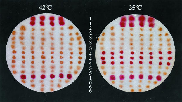
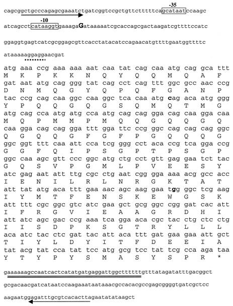
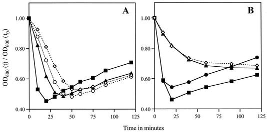
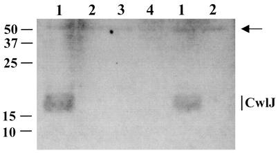
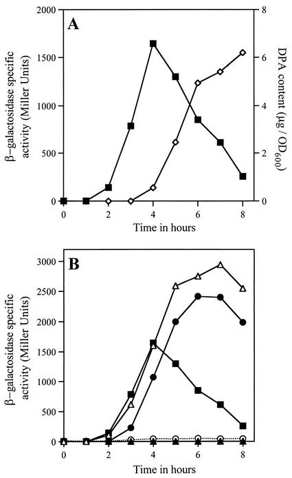
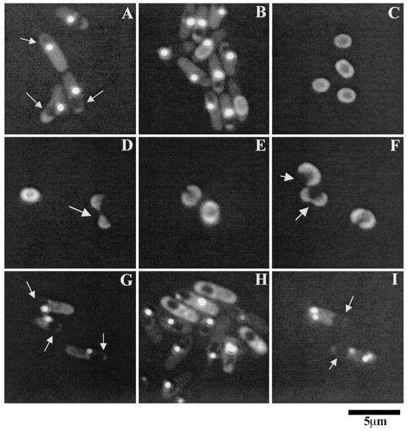
Bacillus subtilis spores can germinate with a 1:1 chelate of Ca(2+) and dipicolinic acid (DPA), a compound present at high levels in the spore core. Using a genetic screen to identify genes encoding proteins that are specifically involved in spore germination by Ca(2+)-DPA, three mutations were identified. One was in the gene encoding the cortex lytic enzyme, CwlJ, that was previously shown to be essential for spore germination by Ca(2+)-DPA. The other two were mapped to an open reading frame, ywdL, encoding a protein of unknown function. Analysis of ywdL expression showed that the gene is expressed during sporulation in the mother cell compartment of the sporulating cell and that its transcription is sigma(E) dependent. Functional characterization of YwdL demonstrated that it is a new spore coat protein that is essential for the presence of CwlJ in the spore coat. Assembly of YwdL itself into the spore coat is dependent on the coat morphogenetic proteins CotE and SpoIVA. However, other than lacking CwlJ, ywdL spores have no obvious defect in their spore coat. Because of the role for YwdL in a part of the spore germination process, we propose renaming ywdL as a spore germination gene, gerQ.


Figures

FIG. 1.
FIG. 1.
Germination of spores with Ca2+-DPA at 42 and 25°C as determined by the plate assay. Well-sporulated colonies were transferred onto a filter paper and incubated with Ca2+-DPA, glucose, and a tetrazolium dye at either 42 or 25°C as described in Materials and Methods. Colonies of germinated spores appear red, due to their ability to carry out metabolism and reduce the tetrazolium dye, while colonies of dormant spores appear brown, because they do not reduce the dye. Horizontal rows of colonies (numbered in the center) are from the following strains: 1, PS832 (wild type); 2, KB1 (mut-5); 3, KB2 (mut-16 ΔyndDEF); 4, KB76 [ger-3b mut-76(Ts)]; 5, KB29 (ΔywdL); and 6, FB111 (ΔcwlJ). Note that ΔcwlJ spores do not germinate with Ca2+-DPA at either temperature.
FIG. 2.
FIG. 2.
Location of mut-5 and mut-16 mutations within the ywdL coding region. The DNA sequence is shown with the predicted YwdL protein sequence in the one-letter code over the first base in each codon. Upstream of the coding region, the −10 and −35 sequences recognized by the sporulation-specific transcription factor σE are shown in boxes. The ywdL transcription start site was determined by 5′ RACE-PCR as described in Materials and Methods and is in an uppercase boldface letter; the putative ribosome binding site is indicated with the broken underline. A putative transcription terminator sequence downstream of the coding region is doubly underlined. The arrows upstream and downstream of the coding region indicate the beginning and the end of the chromosomal region that complemented the mut-5 (KB1) and mut-16 ΔyndDEF (KB2) mutants, as described in Materials and Methods. The sites of the mut-5 [Gln39 (CAG)→stop codon (TAG)] and the mut-16 [Trp123 (TGG)→stop codon (TAG)] mutations are in boldface. The consensus sequence for σE is ATa (16- to 18-bp gap) cATAcaxT, where x can be any nucleotide, uppercase letters indicate highly conserved positions, and lowercase letters indicate less conserved positions (adapted from reference 15).
FIG. 3.
FIG. 3.
Germination of spores in nutrients. (A) Spores of the wild-type strain PS832 (▪) or mutant strain KB1 (ywdL5) (⋄), KB2 (ywdL16) (○), or KB29 (ΔywdL) (▴) were heat activated and incubated in 2× YT medium with 10 mM l-alanine at 37°C. The OD600 of each sample was measured at various time intervals [OD600 (t)] and plotted as the fraction of the initial OD600 at time zero [OD600 (t)/OD600 (t0)] versus time. (B) Spores from wild-type strain PS832 (▪) or mutant strain FB112 (ΔsleB) (•), FB113 (ΔcwlJ ΔsleB) (⋄), or KB30 (ΔsleB ΔywdL) (▴) were germinated in nutrients, and the OD600 of each sample was measured as described above.
FIG. 4.
FIG. 4.
Localization of CwlJ-His tag in spore extracts of various strains. The insoluble fraction of the extract from 1 mg of spores of strains PS3449 (cwlJ-His tag) (lanes 1), KB33 (cwlJ-His tag ΔywdL) (lanes 2), KB29 (ΔywdL) (lane 3), and KB34 (cwlJ-His tag ywdL16) (lane 4) was run on an SDS-12.5% polyacrylamide gel. Each lane contains protein from 1 mg (dry weight) of spores. The gel was blotted onto an Immobilon-P membrane (Millipore) that was hybridized with anti-His tag monoclonal antibody (Novagen) as outlined in Materials and Methods. An extract from spores of strain KB29 (lane 4) that lacks the CwlJ-His tag served as a negative control for the anti-His tag antibody. The arrow indicates an immunoreactive product at ∼50 kDa (with no His tag attached) that has been seen previously (5) and that we use here as a rough indication of protein levels. The CwlJ-His tag runs as a wide band of ∼18 kDa as shown previously (5). Only a portion of the Western blot is shown, and the numbers shown at the left indicate the migration positions of protein molecular mass markers in kilodaltons. The same experiment was run for the soluble fraction of the spore extracts, where CwlJ-His tag was not found (data not shown), as reported previously (5).
FIG. 5.
FIG. 5.
(A) Expression of ywdL-lacZ during sporulation. Strain KB43 (ywdL-lacZ) was induced to sporulate by resuspension at time t0, and samples were removed for estimation of β-galactosidase specific activity (▪) and DPA content (⋄) as described in Materials and Methods. The DPA curve is used here as a marker of the progression through sporulation. (B) Expression of ywdL-lacZ in strains that lack one of the sporulation-specific RNA polymerase σ factors. Strains KB46 (sigF ywdL-lacZ) (○), KB45 (sigE ywdL-lacZ) (▴), KB47 (sigG ywdL-lacZ) (•), KB44 (sigK ywdL-lacZ) (▵), and KB43 (ywdL-lacZ) (▪) were induced to sporulate by resuspension, and the β-galactosidase specific activity was determined as described for panel A. The maximum β-galactosidase specific activity of strain PY79, which was induced to sporulate in parallel with the mutant strains, was found to be 4 Miller units.
FIG. 6.
FIG. 6.
Localization of YwdL-GFP in sporulating cells and dormant spores. (A to C) Strain KB48 (ywdL-gfp), was sporulated by resuspension, and samples were viewed with a fluorescence microscope at 3 h (A), 7 h (B), and 24 h (C) after the onset of sporulation. (D to F) Dormant spores of strain KB48 were germinated in nutrients at 37°C for 1 h, and samples were examined. Arrows indicate germinated spores whose coats have split open. (G to I) Strains KB62 (ΔcotE ywdL-gfp) (G and H) and CVO1736 (ΔspoIVA ywdL-gfp) (I) were sporulated by resuspension, and samples were viewed at 3 h (G and I) and 7 h (H) after the onset of sporulation. Strain KB59 released dormant spores with no visible YwdL-GFP on their periphery, while strain CVO1736 did not release any dormant spores. The arrows pointing to the dark areas enclosed in the sporulating cell in panels A, G, and I indicate the developing forespore.
FIG. 7.
FIG. 7.
Protein sequence alignment of YwdL from various Bacillus species. Asterisks below the aligned sequences indicate identical residues in that column, double dots indicate well conserved residues, and single dots indicate poorly conserved residues (as determined from the ClustalW website). The residues that were altered in strains KB1 and KB2 (Gln39 and Trp123, respectively) are in boldface in the B. subtilis sequence. No homologs of YwdL were found in Clostridium species (15).

Similar articles

Cited by

References

    1. Anagnostopoulos, C., and J. Spizizen. 1961. Requirements for transformation in Bacillus subtilis. J. Bacteriol. 81:74-76. - PMC - PubMed
    1. Atrih, A. P. Z., G. Allmaier, and S. Foster. 1996. Structural analysis of Bacillus subtilis 168 endospore peptidoglycan and its role during differentiation. J. Bacteriol. 178:6173-6183. - PMC - PubMed
    1. Bagyan, I., L. Casillas-Martinez, and P. Setlow. 1998. The katX gene, which codes for the catalase in spores of Bacillus subtilis, is a forespore-specific gene controlled by σF, and KatX is essential for hydrogen peroxide resistance of the germinating spore. J. Bacteriol. 180:2057-2062. - PMC - PubMed
    1. Bagyan, I., B. Setlow, and P. Setlow. 1998. New small, acid-soluble proteins unique to spores of Bacillus subtilis: identification of the coding genes and regulation and function of two of these genes. J. Bacteriol. 180:6704-6712. - PMC - PubMed
    1. Bagyan, I., and P. Setlow. 2002. Localization of the cortex lytic enzyme CwlJ in spores of Bacillus subtilis. J. Bacteriol. 184:1219-1224. - PMC - PubMed

Publication types

MeSH terms

LinkOut - more resources