

Introduction and characterization of a functionally linked metal ion binding site at the exposed heme edge of myoglobin
- PMID: 12644706
- PMCID: PMC152976
- DOI: 10.1073/pnas.0636702100
Introduction and characterization of a functionally linked metal ion binding site at the exposed heme edge of myoglobin
Abstract
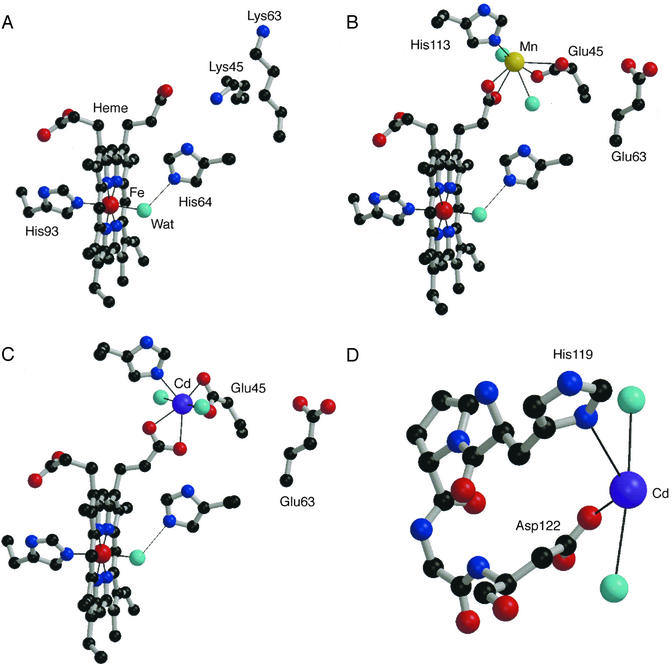
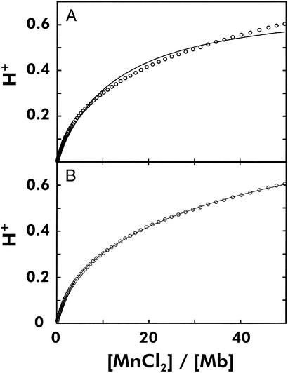
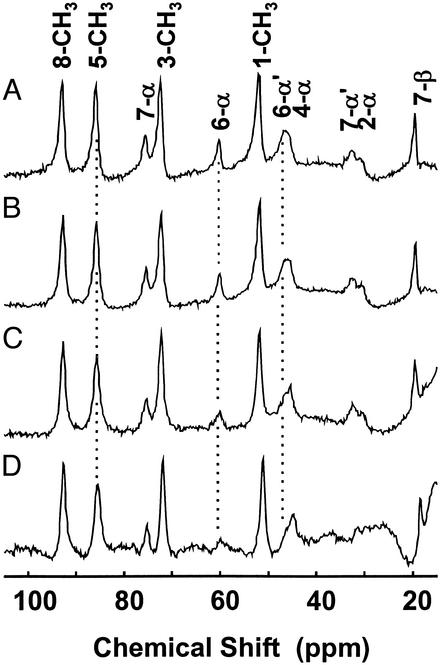
A binding site for metal ions has been created on the surface of horse heart myoglobin (Mb) near the heme 6-propionate group by replacing K45 and K63 with glutamyl residues. One-dimensional (1)H NMR spectroscopy indicates that Mn(2+) binds in the vicinity of the heme 6-propionate as anticipated, and potentiometric titrations establish that the affinity of the new site for Mn(2+) is 1.28(4) x 10(4) M(-1) (pH 6.96, ionic strength I = 17.2 microM, 25 degrees C). In addition, these substitutions lower the reduction potential of the protein and increase the pK(a) for the water molecule coordinated to the heme iron of metmyoglobin. The peroxidase [2,2'-azinobis(3-ethylbenzthiazoline-6-sulfonic acid), ABTS, as substrate] and the Mn(2+)-peroxidase activity of the variant are both increased approximately 3-fold. In contrast to wild-type Mb, both the affinity for azide and the midpoint potential of the variant are significantly influenced by the addition of Mn(2+). The structure of the variant has been determined by x-ray crystallography to define the coordination environment of bound Mn(2+) and Cd(2+). Although slight differences are observed between the geometry of the binding of the two metal ions, both are hexacoordinate, and neither involves coordination by E63.
Figures
Similar articles
-
Electrostatic modification of the active site of myoglobin: characterization of the proximal Ser92Asp variant.Biochemistry. 1996 Sep 10;35(36):11901-12. doi: 10.1021/bi9608976. Biochemistry. 1996. PMID: 8794773
-
Molecular engineering of myoglobin: influence of residue 68 on the rate and the enantioselectivity of oxidation reactions catalyzed by H64D/V68X myoglobin.Biochemistry. 2003 Sep 2;42(34):10174-81. doi: 10.1021/bi034605u. Biochemistry. 2003. PMID: 12939145
-
The stability of holomyoglobin is determined by heme affinity.Biochemistry. 1996 Sep 3;35(35):11310-8. doi: 10.1021/bi9603736. Biochemistry. 1996. PMID: 8784185
-
'It's hollow': the function of pores within myoglobin.J Exp Biol. 2010 Aug 15;213(Pt 16):2748-54. doi: 10.1242/jeb.042994. J Exp Biol. 2010. PMID: 20675544 Review.
-
New functionalization of myoglobin by chemical modification of heme-propionates.Acc Chem Res. 2002 Jan;35(1):35-43. doi: 10.1021/ar000087t. Acc Chem Res. 2002. PMID: 11790087 Review.
Cited by
-
Recycling of the high valence States of heme proteins by cysteine residues of THIMET-oligopeptidase.PLoS One. 2013 Nov 1;8(11):e79102. doi: 10.1371/journal.pone.0079102. eCollection 2013. PLoS One. 2013. PMID: 24223886 Free PMC article.
-
Unravelling the Interactions between Hydrolytic and Oxidative Enzymes in Degradation of Lignocellulosic Biomass by Sporothrix carnis under Various Fermentation Conditions.Biochem Res Int. 2016;2016:1614370. doi: 10.1155/2016/1614370. Epub 2016 Jan 11. Biochem Res Int. 2016. PMID: 26881077 Free PMC article.
-
Kinetic and crystallographic studies of a redesigned manganese-binding site in cytochrome c peroxidase.J Biol Inorg Chem. 2007 Jan;12(1):126-37. doi: 10.1007/s00775-006-0171-0. Epub 2006 Oct 5. J Biol Inorg Chem. 2007. PMID: 17021923
References
Publication types
MeSH terms
Substances
Associated data
- Actions
- Actions
- Actions
- Actions
LinkOut - more resources
Full Text Sources
Miscellaneous
