Comparative Study
. 2003 Apr;72(4):839-49.
doi: 10.1086/373883. Epub 2003 Mar 17.

Missense mutations in the regulatory domain of PKC gamma: a new mechanism for dominant nonepisodic cerebellar ataxia

Affiliations
Comparative Study

Missense mutations in the regulatory domain of PKC gamma: a new mechanism for dominant nonepisodic cerebellar ataxia

Dong-Hui Chen et al. Am J Hum Genet. 2003 Apr.

Abstract

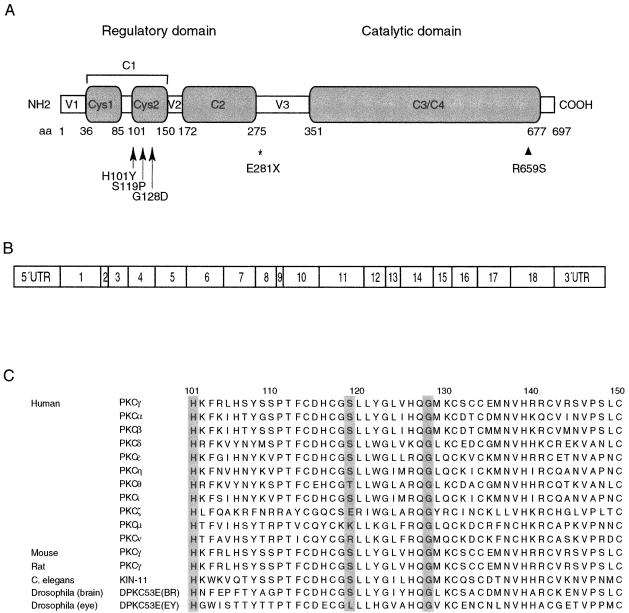
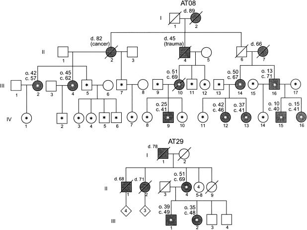
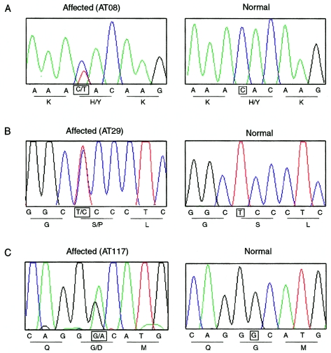
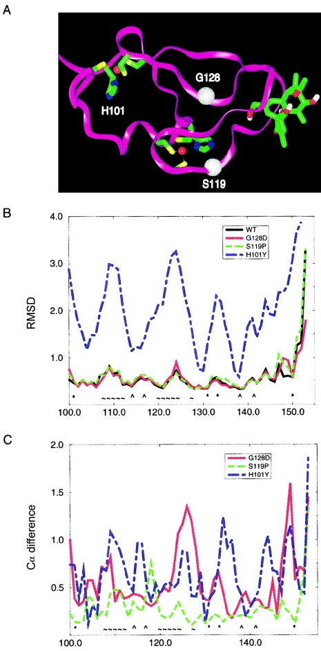
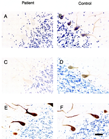
We report a nonepisodic autosomal dominant (AD) spinocerebellar ataxia (SCA) not caused by a nucleotide repeat expansion that is, to our knowledge, the first such SCA. The AD SCAs currently comprise a group of > or =16 genetically distinct neurodegenerative conditions, all characterized by progressive incoordination of gait and limbs and by speech and eye-movement disturbances. Six of the nine SCAs for which the genes are known result from CAG expansions that encode polyglutamine tracts. Noncoding CAG, CTG, and ATTCT expansions are responsible for three other SCAs. Approximately 30% of families with SCA do not have linkage to the known loci. We recently mapped the locus for an AD SCA in a family (AT08) to chromosome 19q13.4-qter. A particularly compelling candidate gene, PRKCG, encodes protein kinase C gamma (PKC gamma), a member of a family of serine/threonine kinases. The entire coding region of PRKCG was sequenced in an affected member of family AT08 and in a group of 39 unrelated patients with ataxia not attributable to trinucleotide expansions. Three different nonconservative missense mutations in highly conserved residues in C1, the cysteine-rich region of the protein, were found in family AT08, another familial case, and a sporadic case. The mutations cosegregated with disease in both families. Structural modeling predicts that two of these amino acid substitutions would severely abrogate the zinc-binding or phorbol ester-binding capabilities of the protein. Immunohistochemical studies on cerebellar tissue from an affected member of family AT08 demonstrated reduced staining for both PKC gamma and ataxin 1 in Purkinje cells, whereas staining for calbindin was preserved. These results strongly support a new mechanism for neuronal cell dysfunction and death in hereditary ataxias and suggest that there may be a common pathway for PKC gamma-related and polyglutamine-related neurodegeneration.


Figures

Figure  1
Figure  1
Organization of PRKCG and corresponding protein. A, Functional domains, showing the conserved (C) and variable (V) regions. The amino acid (aa) boundaries of the regions are shown as predicted in the SMART database. The mutations found in the present study are indicated by arrows. The position of a reported mutation in retinitis pigmentosa (Al-Maghtheh et al. 1998) is indicated by an arrowhead, and the relative position of the rat agu mutation (Craig et al. 2001) is indicated by an asterisk. B, Exonic organization. C, Amino acid sequence alignment in the Cys2 region, showing striking evolutionary conservation of PKCγ and other isozymes of PKC at three mutation sites found in the present study (shown in gray).
Figure  2
Figure  2
Pedigrees of two multiplex kindreds in which PRKCG mutations were identified. Asterisks denote subjects who provided blood samples. Symbols representing clinically affected individuals are blackened, and deceased individuals are indicated by a slash mark. Age at death (d.), approximate age at onset of symptoms (o.), and current age (c.) are shown, in years, above and to the left of the relevant symbols.
Figure  3
Figure  3
Sequence chromatograms for portions of PRKCG exon 4, showing heterozygous mutations in affected individuals from three families with SCA as compared to control individuals. A, A C→T transition in nucleotide 301 in family AT08. B, A T→C transition in nucleotide 355 in family AT29. C, A G→A transition in nucleotide 383 in family AT117. The relevant nucleotides are highlighted by squares. Predicted amino acids are also shown under the corresponding nucleotide sequence.
Figure  4
Figure  4
MD studies of wild-type and mutant Cys2 regions of PKCγ. A,Three-dimensional structure of the Cys2 region of wild-type PKCγ with phorbol acetate bound. H101 is a ligand in the first zinc binding site; zinc is shown in red. S119 is close to the second zinc binding site, as well as the phorbol acetate binding site. G128 is also in the vicinity of the phorbol acetate binding site; Cα positions of S119 and G128 are indicated by white spheres. B, Fluctuations of protein backbone coordinates per residue, expressed as root-mean-square deviation (RMSD) (in Å) for wild type and three mutants. Note the instability of the H101Y mutant in comparison to wild type and the two other mutants. C, Differences in average structures, calculated as distance between Cα positions (in Å) per residue between wild type and each of the three mutants. Note the global shift of the structure in the H101Y mutant and the shifts in the phorbol ester binding site in the G128D mutant. WT = wild type; * = first zinc binding site; ⁁ = second zinc binding site; ∼ = phorbol binding site.
Figure  5
Figure  5
Reduced immunoreactivity for PKCγ and ataxin 1 in an ataxic patient from family AT08 who carries the H101Y mutation. A and B, PKCγ immunohistochemistry. The scattered surviving Purkinje cells in a postmortem cerebellar section showed reduced but variable staining for antibody to PKCγ (A), compared with intense staining in membrane and dendrites from an age-matched control (B). C and D, Ataxin 1 immunohistochemistry. The majority of Purkinje cells from the same ataxic patient showed no immunostaining for ataxin 1, but a minority of cells were weakly positive. Control Purkinje cells were intensely stained in cytoplasm (D). E and F, Calbindin immunohistochemistry. Staining for calbindin was similar in the ataxic patient and the control individual, demonstrating that the loss of PKCγ and ataxin 1 did not represent nonspecific decreased protein synthesis. Scale bar = 75 μm.

References

Electronic-Database Information

    1. Entrez Genome View, http://www.ncbi.nlm.nih.gov/mapview/map_search.cgi?chr=hum_chr.inf (for NCBI Map Viewer)
    1. GenBank, http://www.ncbi.nlm.nih.gov/Genbank/ (for PRKCG mRNA sequence [accession number NM_002739])
    1. GeneTests Home Page, http://www.geneclinics.org/ or http://www.genetests.org/ (for “Hereditary Ataxia Overview,” by T.D.B., in GeneReviews)
    1. Online Mendelian Inheritance in Man (OMIM), http://www.ncbi.nlm.nih.gov/Omim/ (for PRKCG) - PubMed
    1. SMART, http://smart.embl-heidelberg.de/ (for domain prediction within PKCγ)

References

    1. Abeliovich A, Paylor R, Chen C, Kim JJ, Wehner JM, Tonegawa S (1993) PKC γ mutant mice exhibit mild deficits in spatial and contextual learning. Cell 75:1263–1271 - PubMed
    1. Al-Maghtheh M, Vithana EN, Inglehearn CF, Moore T, Bird AC, Bhattacharya SS (1998) Segregation of a PRKCG mutation in two RP11 families. Am J Hum Genet 62:1248–1252 - PMC - PubMed
    1. Barmack NH, Qian Z, Yoshimura J (2000) Regional and cellular distribution of protein kinase C in rat cerebellar Purkinje cells. J Comp Neurol 427:235–254 - PubMed
    1. Brkanac Z, Bylenok L, Fernandez M, Matsushita M, Lipe H, Wolff, J, Nochlin D, Raskind WH, Bird TD (2002) A new dominant spinocerebellar ataxia is linked to chromosome 19q13.4. Arch Neurol 59:1291–1295 - PubMed
    1. Burright EN, Clark HB, Servadio A, Matilla T, Feddersen RM, Yunis WS, Duvick LA, Zoghbi HY, Orr HT (1995) SCA1 transgenic mice: a model for neurodegeneration caused by an expanded CAG trinucleotide repeat. Cell 82:937–948 - PubMed

Publication types

Associated data