

Rat testicular germ cells and Sertoli cells release different types of bioactive transforming growth factor beta in vitro
- PMID: 12646048
- PMCID: PMC151560
- DOI: 10.1186/1477-7827-1-3
Rat testicular germ cells and Sertoli cells release different types of bioactive transforming growth factor beta in vitro
Abstract
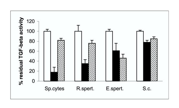
Several in vivo studies have reported the presence of immunoreactive transforming growth factor-beta's (TGF-beta's) in testicular cells at defined stages of their differentiation. The most pronounced changes in TGF-beta1 and TGF-beta2 immunoreactivity occurred during spermatogenesis. In the present study we have investigated whether germ cells and Sertoli cells are able to secrete bioactive TGF-beta's in vitro, using the CCl64 mink lung epithelial cell line as bioassay for the measurement of TGF-beta. In cellular lysates, TGF-beta bioactivity was only observed following heat-treatment, indicating that within these cells TGF-beta is present in a latent form. To our surprise, active TGF-beta could be detected in the culture supernatant of germ cells and Sertoli cells without prior heat-treatment. This suggests that these cells not only produce and release TGF-beta in a latent form, but that they also release a factor which can convert latent TGF-beta into its active form. Following heat-activation of these culture supernatant's, total TGF-beta bioactivity increased 6- to 9-fold. Spermatocytes are the cell type that releases most bioactive TGF-beta during a 24 h culture period, although round and elongated spermatids and Sertoli cells also secrete significant amounts of TGF-beta. The biological activity of TGF-beta could be inhibited by neutralizing antibodies against TGF-beta1 (spermatocytes and round spermatids) and TGF-beta2 (round and elongating spermatids). TGF-beta activity in the Sertoli cell culture supernatant was inhibited slightly by either the TGF-beta1 and TGF-beta2 neutralizing antibody. These in vitro data suggest that germ cells and Sertoli cells release latent TGF-beta's. Following secretion, the TGF-beta's are converted to a biological active form that can interact with specific TGF-beta receptors. These results strengthen the hypothesis that TGF-beta's may play a physiological role in germ cell proliferation/differentiation and Sertoli cell function.
Figures
Similar articles
-
Localization of transforming growth factor beta 1 and beta 2 during testicular development in the rat.Biol Reprod. 1993 Jan;48(1):40-5. doi: 10.1095/biolreprod48.1.40. Biol Reprod. 1993. PMID: 8418916
-
Basic fibroblast growth factor is a testicular germ cell product which may regulate Sertoli cell function.Mol Endocrinol. 1993 Jul;7(7):889-97. doi: 10.1210/mend.7.7.8413313. Mol Endocrinol. 1993. PMID: 8413313
-
Sertoli cell-germ cell interactions and TGF beta 1 expression and secretion in vitro.Biochem Biophys Res Commun. 1997 Sep 29;238(3):905-9. doi: 10.1006/bbrc.1997.7275. Biochem Biophys Res Commun. 1997. PMID: 9325190
-
Regulation of aromatase gene expression in Leydig cells and germ cells.J Steroid Biochem Mol Biol. 2003 Sep;86(3-5):335-43. doi: 10.1016/s0960-0760(03)00343-1. J Steroid Biochem Mol Biol. 2003. PMID: 14623530 Review.
-
Interactions between FSH, estradiol-17 beta and transforming growth factor-beta regulate growth and differentiation in the rat gonad.J Steroid Biochem Mol Biol. 1993 Mar;44(4-6):441-7. doi: 10.1016/0960-0760(93)90248-u. J Steroid Biochem Mol Biol. 1993. PMID: 8476758 Review.
Cited by
-
Eosinophil cationic protein stimulates TGF-beta1 release by human lung fibroblasts in vitro.Inflammation. 2007 Oct;30(5):153-60. doi: 10.1007/s10753-007-9032-4. Inflammation. 2007. PMID: 17587163
-
Transforming growth factor beta-1 decreases the yield of the second meiotic division of rat pachytene spermatocytes in vitro.Reprod Biol Endocrinol. 2005 Jun 7;3:22. doi: 10.1186/1477-7827-3-22. Reprod Biol Endocrinol. 2005. PMID: 15941479 Free PMC article.
-
Fractionated irradiation of right thorax induces abscopal damage on testes leading to decline in fertility.Sci Rep. 2019 Oct 23;9(1):15221. doi: 10.1038/s41598-019-51772-y. Sci Rep. 2019. PMID: 31645625 Free PMC article.
References
-
- Bellvé AR, Zheng W. Growth factors as autocrine and paracrine modulators of male gonadale functions. J Reprod Fertil. 1989;85:771–793. - PubMed
-
- Avallet O, Vigier M, Albaladejo V, de Cesaris P, Saez JM. Transforming growth factor β gene expression in cultured porcine Sertoli and Leydig cells: Effects of hormine and growth factors. Proceedings of the 6th European Workshop on Molecular and Cellular Endocrinology of the Testis. 1990;D13
-
- Caussanel S, Tabone E, Hendrick JC, Dacheux F, Benahmed M. Cellular distribution of transforming growth factor betas 1, 2, and 3 and their types I and II receptors during postnatal development and spermatogenesis in the boar testis. Biol Reprod. 1997;56:357–367. - PubMed
-
- Watrin F, Scotto L, Associan RK, Wolgemuth DJ. Cell lineage specificity of expression of the murine transforming growth factor-β3 and transforming growth factor-β1 genes. Cell Growth & Differ. 1991;2:77–83. - PubMed
-
- Skinner MK, Moses HL. Transforming growth factorβ gene expression and action in the seminiferous tubule: Peritubular cell-Sertoli cell interactions. Mol Endocrinol. 1989;3:625–634. - PubMed
Publication types
MeSH terms
Substances
LinkOut - more resources
Full Text Sources
