. 2003 Apr 1;412(1):106-20.
doi: 10.1016/s0003-9861(03)00012-2.

Expression and purification of recombinant tristetraprolin that can bind to tumor necrosis factor-alpha mRNA and serve as a substrate for mitogen-activated protein kinases

Affiliations

Expression and purification of recombinant tristetraprolin that can bind to tumor necrosis factor-alpha mRNA and serve as a substrate for mitogen-activated protein kinases

Heping Cao et al. Arch Biochem Biophys. .

Abstract

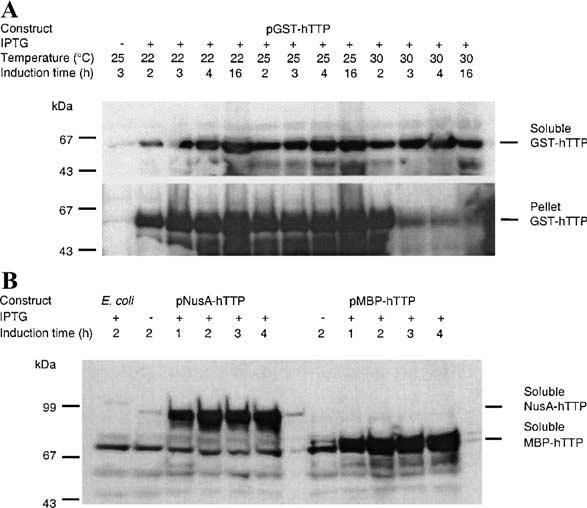
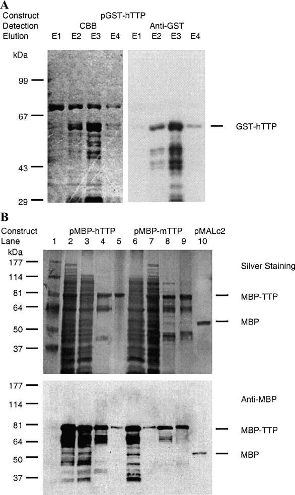
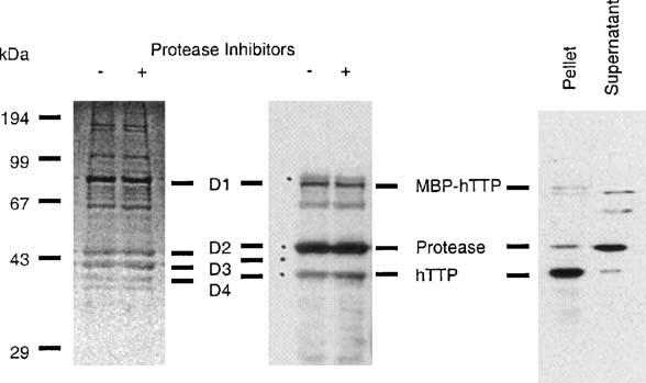
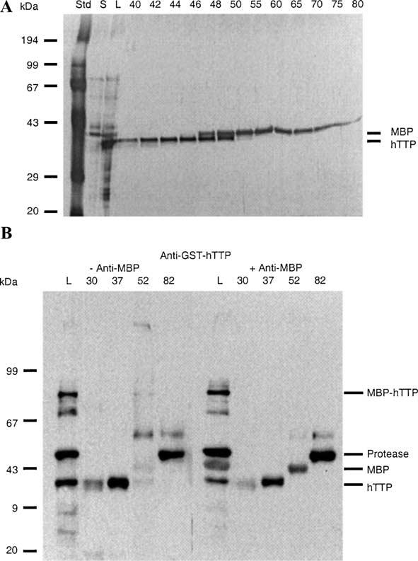
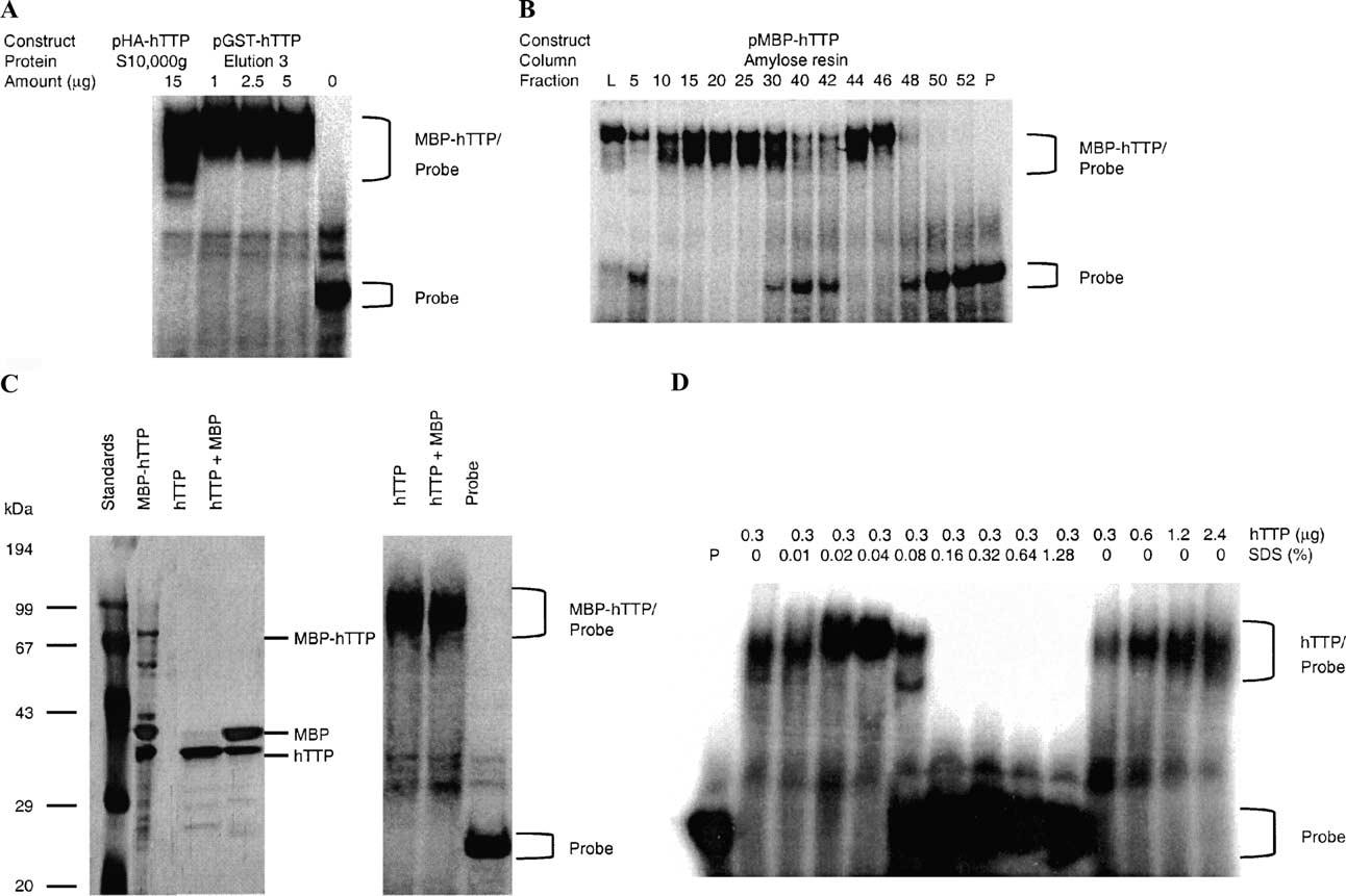
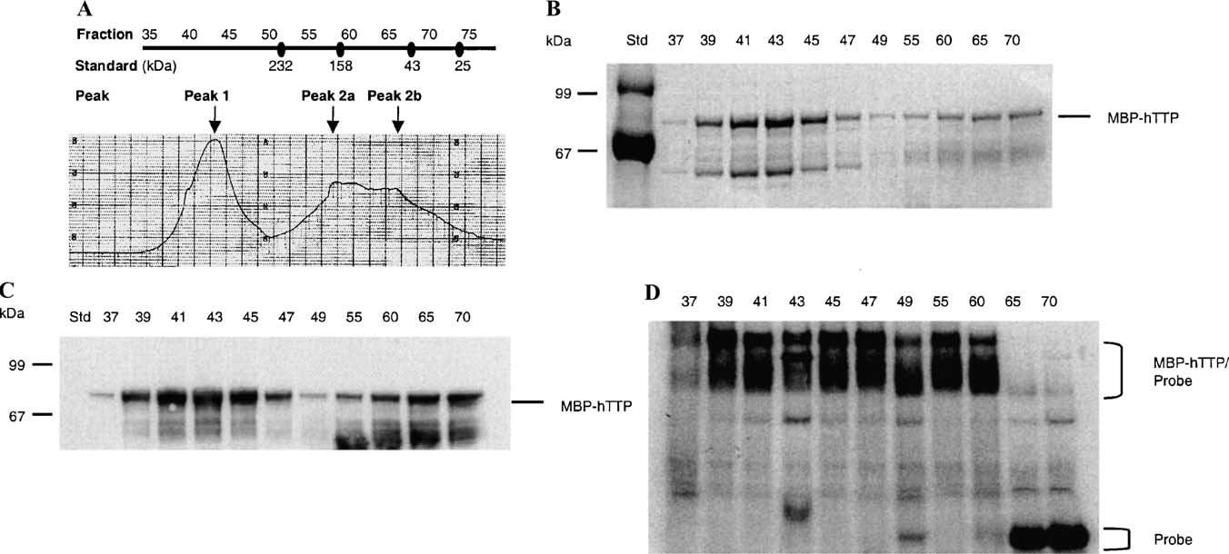
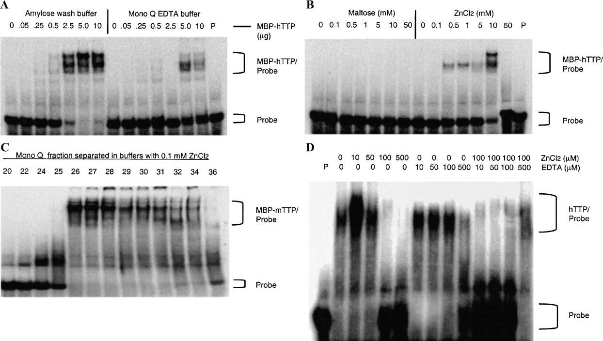
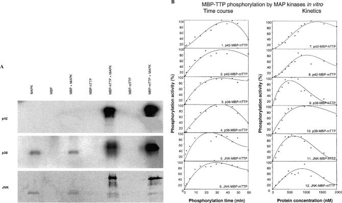
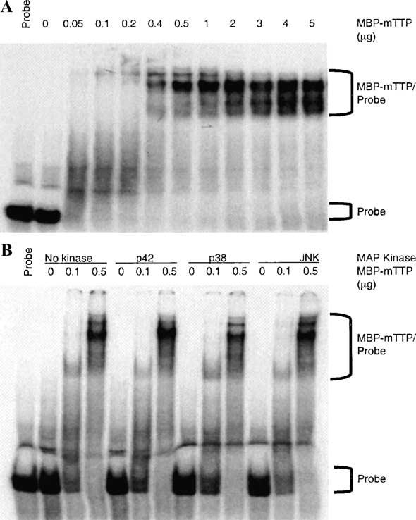
Tristetraprolin (TTP) is an mRNA-binding protein, but studies of this interaction have been difficult due to problems with the purification of recombinant TTP. In the present study, we expressed human and mouse TTP as glutathione S-transferase and maltose-binding protein (MBP) fusion proteins in Escherichia coli, and purified them by affinity resins and Mono Q chromatography. TTP cleaved from the fusion protein was identified by immunoblotting, MALDI-MS, and protein sequencing, and was further purified to homogeneity by continuous-elution SDS-gel electrophoresis. Purified recombinant TTP bound to the AU-rich element of tumor necrosis factor-alpha (TNFalpha) mRNA and this binding was dependent on Zn(2+). Results from sizing columns suggested that the active species might be in the form of an oligomer of MBP-TTP. Recombinant TTP was phosphorylated by three members of the mitogen-activated protein (MAP) kinase family, p42, p38, and JNK, with half-maximal phosphorylation occurring at approximately 0.5, 0.25, and 0.25 microM protein, respectively. Phosphorylation by these kinases did not appear to affect the ability of TTP to bind to TNFalpha mRNA under the assay conditions. This study describes a procedure for purifying nonfusion protein TTP to homogeneity, demonstrates that TTP's RNA-binding activity is zinc dependent, and that TTP can be phosphorylated by JNK as well as by the other members of the greater MAP kinase family.


Figures

Fig. 1
Fig. 1
Expression of recombinant hTTP in E. coli. E. coli BL21(DE3) cells were transformed with expression plasmids pGST-hTTP, pMBP-hTTP, and pNusA-hTTP. The fusion proteins were induced by 0.1, 0.4, and 0.4 mM IPTG, respectively, at 22, 25, 30 °C (for pGST-hTTP), and 28 °C (for pMBP-hTTP and pNusA-hTTP) for various times as indicated. (A) GST-hTTP in the 10,000g supernatant and the pellet from the same volume of homogenate were analyzed by immunoblotting using anti-GST serum. Immunoreactive GST-hTTP is indicated. (B) MBP-hTTP and NusA-hTTP in the 10,000g supernatant were detected by immunoblotting using affinity-purified anti-GST-hTTP, as indicated. Similar results were observed using plasmid pMBP-mTTP (data not shown). Lanes labeled E. coli and no IPTG(−) as two negative controls were soluble extracts from E. coli cells without plasmids and E. coli cells with plasmids but without IPTG induction, respectively.
Fig. 2
Fig. 2
Purification of recombinant TTP fusion proteins and MBP from E. coli. GST-hTTP, MBP-hTTP, MBP-mTTP, and MBP were induced by IPTG in E. coli as described in the legend for Fig. 1. (A) GST-hTTP was affinity-purified with glutathione Sepharose 4B beads and eluted from the beads four times successively, designating elutions 1, 2, 3, and 4 as E1, E2, E3, and E4. The eluted proteins were separated with 8% SDS–PAGE and detected with Commassie brilliant blue (CBB) staining (left panel) or immunoblotting with anti-GST serum (right panel). Immunoreactive GST-hTTP is indicated. (B) MBP-hTTP, MBP-mTTP, and MBP were initially affinity-purified from the 10,000g supernatant by an amylose resin affinity column and eluted with 10 mM maltose. MBP-hTTP (left panel) and MBP-mTTP (right panel) from the amylose resin column were further purified using a Mono Q column. Top panel: 10% SDS–PAGE separation and silver staining. Lane 1, protein molecular size standards; lane 2, total E. coli homogenate expressing MBP-hTTP (50 μg of protein); lane 3, S10,000g supernatant (50 μg); lane 4, peak fraction eluted from the amylose resin column (5 μg); lane 5, peak fraction eluted from the Mono Q column (1 μg). Lane 6, total E. coli homogenate expressing MBP-mTTP (50 μg); lane 7, S10,000g supernatant (50 μg); lane 8, peak fraction eluted from the amylose resin column (5 μg); lane 9, peak fraction eluted from the Mono Q column (5 μg); and lane 10, the peak fraction of MBP from amylose resin column (5 μg). The positions of purified MBP-TTP (both hTTP and mTTP) and MBP are indicated. Bottom panel: Immunological detection of MBP-hTTP, MBP-mTTP, and MBP by anti-MBP serum. The samples were identical to those used in the upper panel except that 10% of the amount of protein used in the upper panel was used.
Fig. 3
Fig. 3
PreScission protease digestion of MBP-hTTP and precipitation of hTTP following the digestion. Left panel: MBP-hTTP from the amylose resin column was digested at 5 °C for 18 h in the digestion buffer with or without 1 mM PMSF and 2 μM leupeptin (protease inhibitors), separated by 12% SDS–PAGE, and stained with CBB. Middle panel: Proteins were digested, separated by 12% SDS–PAGE, transferred onto nitrocellulose membranes, and detected with anti-GST-hTTP antibodies. The four major protein bands (D1–D4) detected by Ponceau S staining before immunoblotting are indicated. The bands corresponding to MBP-hTTP, the PreScission protease, and hTTP are indicated. Right panel: MBP-hTTP was purified by amylose resin affinity chromatography and digested with PreScission protease overnight. The digestion mixture was centrifuged at 20,000g for 5 min. Proteins in the supernatant and pellet fractions were separated by 12% SDS–PAGE and immunoblotted with anti-GST-hTTP antibodies. The bands corresponding to MBP-hTTP, the PreScission protease, and hTTP are indicated.
Fig. 4
Fig. 4
Purification of hTTP by continuous-elution gel electrophoresis following PreScission protease digestion. Proteins (2 mg in 2 ml) from an overnight digestion as described in the legend of Fig. 3 were separated by 12% SDS–PAGE and eluted continuously with SDS–PAGE running buffer at 1 ml/min. Eluted proteins were collected in 5-ml fractions. (A) Proteins in the digestion mixture (L; 1 μl) and selected fractions between 40 and 80 (20 μl) were separated by 12% SDS–PAGE and visualized by silver staining. The positions of purified hTTP and MBP are indicated. S, the 10,000g supernatant of the protease digestion mixture. (B) Similar fractions from another experiment were concentrated and proteins (1/3 of the concentrated protein per lane) were used for immunoblotting with anti-GST-hTTP antibodies (left panel) or a mixture of anti-GST-hTTP plus anti-MBP antibodies (right panel). Note that the anti-GST-hTTP antibodies recognized both the bottom hTTP band and the top GST fusion protease band whereas the anti-MBP antibody recognized the middle MBP band. L, the protease digestion mixture.
Fig. 5
Fig. 5
ARE-binding activity of GST-hTTP, MBP-hTTP, and hTTP. (A) GST-hTTP-binding activity. Various amounts (0, 1, 2.5, and 5 μg) of GST-hTTP in the third elution from glutathione-Sepharose 4B beads (E3 in Fig. 2A) were used for TNFα mRNA ARE-binding activity assays. As a comparison, HA-hTTP (15 μg of total protein in the 10,000g supernatant) from transfected human HEK 293 cells was used. (B) MBP-hTTP-binding activity. The ARE-binding activity of MBP-hTTP was determined using the TNFα mRNA ARE probe and 1 μl of amylose resin fractions 5 to 25 (unbound), fractions 30 to 42 (wash), and fractions 44 to 52 (elution). L, load (the 10,000g supernatant); P, probe control. Similar results were obtained using MBP-mTTP (data not shown). No activity was observed with fractions from E. coli cells transformed with pMALc2 (gel not shown). (C) Purified hTTP-binding activity. Human TTP was cleaved from MBP-hTTP by PreScission protease and separated from other proteins by CEGE. The CEGE-purified hTTP and the digested MBP-hTTP were separated by 10% SDS–PAGE, silver-stained (left panel), and used in the ARE-binding assay using the TNFα mRNA probe (right panel). (D) Effect of SDS on hTTP's ARE-binding activity. Human TTP purified by CEGE was used for ARE-binding activity assay in buffers containing various amounts of hTTP without SDS (right panel) or various concentrations of SDS with the same amount of hTTP (left panel) as indicated. P, probe control.
Fig. 6
Fig. 6
ARE-binding activity of MBP-hTTP peaks from gel-filtration chromatography. (A) Chromatogram. MBP-hTTP (50 mg) was partially purified by amylose resin chromatography and applied to a Sephacryl S200 column (2:5 × 112 cm). Fraction numbers are indicated on the top. The protein peaks (at 280 nm) were labeled as Peak 1, Peak 2a, and Peak 2b. The approximate molecular masses of proteins were determined using a standard curve generated on the same column. The standard protein sizes are indicated below the fraction numbers and include bovine pancreas ribonuclease A (13.7 kDa), bovine pancreas chymotrypsinogen (25 kDa), hen egg ovalbumin (43 kDa), bovine serum albumin (67 kDa), rabbit muscle aldolase (158 kDa), and bovine liver catalase (232 kDa). (B) SDS–PAGE. Selected protein fractions from the column were separated with 10% SDS–PAGE and stained with CBB. The position of MBP-hTTP is indicated. (C) Immunoblotting. MBP-hTTP in the column fractions was identified by immunoblotting using anti-MBP serum. (D) ARE-binding activity. ARE-binding activity of the column fractions indicated was assayed using the TNFa mRNA probe.
Fig. 7
Fig. 7
Effect of zinc on the ARE-binding activity of MBP-TTP and hTTP. (A) Effect of EDTA on the ARE-binding activity of MBP-TTP. ARE-binding activity was determined using various amounts of MBP-hTTP (as indicated in the figure) eluted from the amylose resin column and diluted with either amylose wash buffer with 10 mM maltose or Mono Q buffer A with 5 mM EDTA. P, probe alone. (B) Effect of zinc on the ARE-binding activity of inactive MBP-TTP. The ARE-binding activity was determined by using the same amount of inactive MBP-hTTP (10 μl) from the Mono Q fractions in Mono Q buffer that were supplemented with various amounts of maltose or ZnCl2, as indicated. P, probe alone. (C) Purification of active MBP-TTP by Mono Q column with zinc-containing buffer. MBP-mTTP from amylose resin was separated by Mono Q column in buffers containing 0.1 mM ZnCl2 and no EDTA. MBP-mTTP ARE-binding activity was determined with GMSA using the TNFα mRNA ARE probe. (D) Effect of zinc and EDTA on ARE-binding activity of hTTP. CEGE-purified hTTP (0.3 μg) was used for ARE-binding activity assay in buffers containing various concentrations of ZnCl2, EDTA, and mixtures of ZnCl2 and EDTA as indicated. P, probe control.
Fig. 8
Fig. 8
Phosphorylation of MBP-TTP (both hTTP and mTTP) by MAP kinases. (A) MBP-TTP (1 μg) was partially purified by amylose resin column and used as a substrate for p42 (0.1 μg), p38 (0.5 μg), or JNK (0.2 μg). The reactions in 20 μl were performed at 30 °C for 30 min. (B) Time course (left panel, plots 1–6) and substrate concentration dependence (right panel, plots 7–12) of the phosphorylation reactions. The reactions were performed at 30 °C for various times using 1 μM MBP-TTP as the substrate in 150 μl (10 μl per time point) (left panel), or at 30 °C for 15 min using various amounts of MBP-TTP in 20 μl (right panel). The results were quantified by Phosphorimager, analyzed by ImageQuant 5.1, and plotted by SigmaPlot 8.0. The data presented show the results of single determinations for plots 1–4 and 9, the means of results of two determinations for plots 5–7, 11, and 12, and the means of results of four determinations for plots 8 and 10. The standard deviations for the means were within 20% of the means. The MBP-TTP concentration was estimated by designating the purity of the MBP-TTP purified from the amylose resin affinity column (Fig. 2B, lanes 4 and 8) as 70%. The ATP used in the reactions for p42, p38, and JNK was [γc-32P]ATP, [γ-32P]ATP, and [γ-33P]ATP, respectively.
Fig. 9
Fig. 9
Effect of phosphorylation of MBP-mTTP by MAP kinases on its ARE-binding activity. MBP-mTTP partially purified from the amylose resin column was used for in vitro phosphorylation and GMSA. (A) The binding activity of various amounts of MBP-mTTP is shown. (B) Based on the results in A, two concentrations of MBP-mTTP and the control without MBP-mTTP were selected for phosphorylation by p42, p38, or JNK kinases, under the same conditions as described in Fig. 8. The reaction mixtures were then used for ARE-binding assays under conditions identical to those used in A.

Similar articles

Cited by

References

    1. DuBois RN, McLane MW, Ryder K, Lau LF, Nathans D. J. Biol. Chem. 1990;265:19185–19191. - PubMed
    1. Lai WS, Stumpo DJ, Blackshear PJ. J. Biol. Chem. 1990;265:16556–16563. - PubMed
    1. Varnum BC, Ma QF, Chi TH, Fletcher B, Herschman HR. Mol. Cell. Biol. 1991;11:1754–1758. - PMC - PubMed
    1. Lai WS, Carballo E, Thorn JM, Kennington EA, Blackshear PJ. J. Biol. Chem. 2000;275:17827–17837. - PubMed
    1. Carballo E, Lai WS, Blackshear PJ. Science. 1998;281:1001–1005. - PubMed

Publication types

MeSH terms