. 2003 Apr 1;100(7):4072-7.
doi: 10.1073/pnas.2628038100. Epub 2003 Mar 19.

Designing isoform-specific peptide disruptors of protein kinase A localization

Affiliations

Designing isoform-specific peptide disruptors of protein kinase A localization

Lora L Burns-Hamuro et al. Proc Natl Acad Sci U S A. .

Abstract

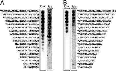
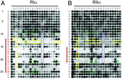
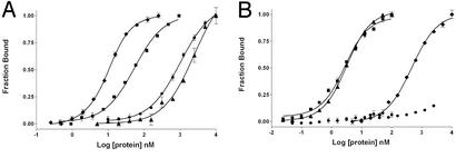
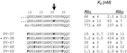
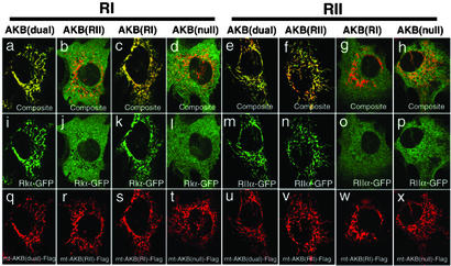
A kinase-anchoring proteins (AKAPs) coordinate cAMP-mediated signaling by binding and localizing cAMP-dependent protein kinase (PKA), using an amphipathic helical docking motif. Peptide disruptors of PKA localization that mimic this helix have been used successfully to assess the involvement of PKA in specific signaling pathways. However, these peptides were developed as disruptors for the type II regulatory subunit (RII) even though both RI and RII isoforms can bind to AKAPs and have discrete functions. To evaluate the effects of each localized isoform, we designed peptides that specifically bind to either RI or RII. Using a peptide array, we have defined the minimal binding sequence of dual specific-AKAP 2 (d-AKAP2), which binds tightly to both RI and RII. Side-chain requirements for affinity and isoform specificity were evaluated by using a peptide substitution array where each position along the A kinase binding domain of d-AKAP2 was substituted by the other 19 l-amino acids. This array comprises 513 single-site substitution analogs of the d-AKAP2 sequence. Peptides containing single and multiple mutations were evaluated in a quantitative fluorescence binding assay and a cell-based colocalization assay. This strategy has allowed us to design peptides with high affinity (K(D) = 1-2 nM) and high specificity for RIalpha versus RIIalpha. These isoform-specific peptides will be invaluable tools to evaluate functional differences between localized RI and RII PKA and are RIalpha-specific disruptors. This array-based analysis also provides a foundation for biophysical analysis of this docking motif.


Figures

Figure 1
Figure 1
N-terminal (A) and C-terminal (B) truncations of the 27-residue AKB domain of D-AKAP2. Truncated peptides were synthesized by using SPOT synthesis on cellulose membrane as described in Materials and Methods. Binding was evaluated by incubating each membrane with GFP-RIα D/D domain and GFP-RIIα D/D domain as indicated. Bound protein was detected by using a primary antibody against GFP and enzyme-conjugated secondary antibody for amplification of signal. The membrane was then analyzed by chemiluminescence.
Figure 2
Figure 2
Peptide substitution array of the 27-residue AKB domain of d-AKAP2 prepared by SPOT synthesis. All 20 amino acids (top of blot) were substituted into each position along the AKB domain (left side of blot). The sequences corresponding to the left column of each array are identical and represent the unsubstituted peptide. Other spots are single substitution analogs. Binding was evaluated by using a conjugated antibody system for both RIα (A) and RIIα (B). The residues highlighted in red show a decreased tolerance for substitutions at these positions. The red bar to the left of each blot highlights this region. Yellow boxes indicate those residues that disrupt binding to RIα while maintaining binding to RIIα. Green circles indicate those positions that enhance binding to RIα while disrupting binding to RIIα. Highlighted in blue are substituted proline residues.
Figure 3
Figure 3
Binding of AKB(dual) (■), AKB(RI) (Q9F, V21W, M25F) (⧫), AKB(RII) (A13L) (▴), and AKB(null) (●) peptides to full-length RIα (A) and RIIα (B). Each peptide was fluorescently labeled and incubated with the corresponding regulatory subunit for 1 h in 10 mM Hepes, 150 mM NaCl, 3 mM EDTA, pH 7.4. Fluorescence anisotropy was used to monitor bound peptide.
Figure 4
Figure 4
Binding dissociation constants (KD) of selected mutant peptides of d-AKAP2. Binding was evaluated for both RIα and RIIα by using fluorescence anisotropy as described in Fig. 3. Substituted residues are bold. Tryptophan at position 21, highlighted with the arrow, is important for discriminating against binding to the RIIα subunit. PV-38 is designated the RI-specific binding peptide, AKB(RI) and is indicated by an asterisk.
Figure 5
Figure 5
Isotype-specific mutations of AKB domain interact with RI or RII distinctively. The mitochondrial-targeting constructs containing WT AKB (q and u), RII-specific mutation (r and v), RI-specific mutation (s and w), or null mutation (t and x) were cotransfected with RIα-GFP (il) or RIIα-GFP (mp), respectively. The interactions between AKB and RI or RII are reflected by the localization of the GFP tag on mitochondria (i, k, m, and n), whereas the noninteracting combinations give a diffused pattern (j, l, o, and p). (ah) The composite images of AKB and R in the same cells.

References

    1. Taylor S S, Buechler J A, Yonemoto W. Annu Rev Biochem. 1990;59:971–1005. - PubMed
    1. Colledge M, Scott J D. Trends Cell Biol. 1999;9:216–221. - PubMed
    1. Doskeland S O, Maronde E, Gjertsen B T. Biochim Biophys Acta. 1993;1178:249–258. - PubMed
    1. Feliciello A, Gottesman M E, Avvedimento E V. J Mol Biol. 2001;308:99–114. - PubMed
    1. Skalhegg B S, Tasken K. Front Biosci. 2000;5:D678–D693. - PubMed

Publication types

MeSH terms

LinkOut - more resources