. 2003 Apr 1;100(7):4120-5.
doi: 10.1073/pnas.0730640100. Epub 2003 Mar 19.

Transforming growth factor beta 1 inhibits expression of NKp30 and NKG2D receptors: consequences for the NK-mediated killing of dendritic cells

Affiliations

Transforming growth factor beta 1 inhibits expression of NKp30 and NKG2D receptors: consequences for the NK-mediated killing of dendritic cells

Roberta Castriconi et al. Proc Natl Acad Sci U S A. .

Abstract

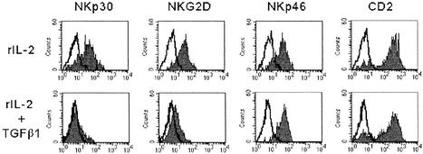
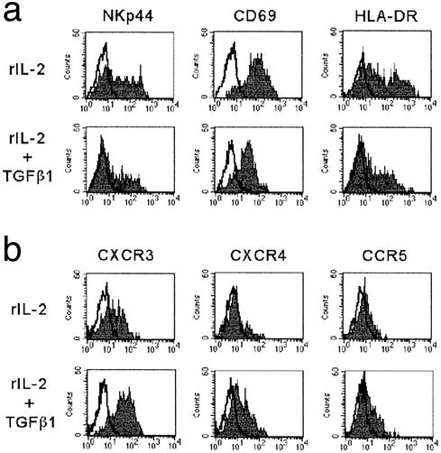
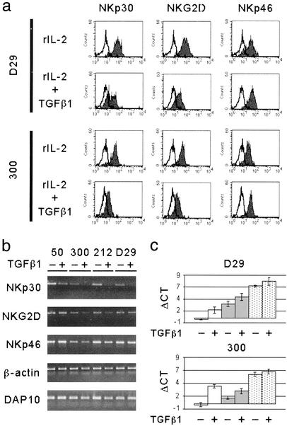
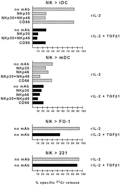
The surface density of the triggering receptors responsible for the natural killer (NK)-mediated cytotoxicity is crucial for the ability of NK cells to kill susceptible target cells. In this study, we show that transforming growth factor beta1 (TGFbeta1) down-regulates the surface expression of NKp30 and in part of NKG2D but not that of other triggering receptors such as NKp46. The TGFbeta1-mediated inhibition of NKp30 surface expression reflects gene regulation at the transcriptional level. NKp30 has been shown to represent the major receptor involved in the NK-mediated killing of dendritic cells. Accordingly, the TGFbeta1-dependent down-regulation of NKp30 expression profoundly inhibited the NK-mediated killing of dendritic cells. On the contrary, killing of different NK-susceptible tumor cell lines was variably affected, reflecting the differential usage of NKp30 and/or NKG2D in the lysis of such tumors. Our present data suggest a possible mechanism by which TGFbeta1-producing dendritic cells may acquire resistance to the NK-mediated attack.


Figures

Figure 1
Figure 1
TGFβ1-mediated down-regulation of NKp30 and NKG2D surface molecules in fresh NK cells. Freshly isolated NK cells were cultured either in the presence of rIL-2 alone or in the presence of rIL-2 and TGFβ1. At day 7 cells were analyzed by one-color immunofluorescence and fluorescence-activated cell sorter analysis for the expression of the indicated molecules. PE-conjugated isotype-specific goat anti-mouse IgG was used as second reagent. White profiles represent cells stained with the second reagent alone.
Figure 2
Figure 2
Expression of activation surface markers and chemokine receptors in TGFβ1-conditioned NK cells. Freshly isolated NK cells were plated either in the presence of rIL-2 alone or in the presence of rIL-2 and TGFβ1. At day 7 cells were analyzed by one-color immunofluorescence and fluorescence-activated cell sorter analysis for the expression of NKp44, CD69, and HLA-DR (a) or of various chemokine receptors (b). PE-conjugated isotype-specific goat anti-mouse IgG was used as second reagent. White profiles represent cells stained with the second reagent alone.
Figure 3
Figure 3
TGFβ1 effect on NKp30 and NKG2D surface and transcript expression in established NK cell population and clones. (a) Long-term rIL-2 cultured polyclonal or clonal NK cells (see the representative polyclonal NK population D29 and NK cell clone 300) were replated in rIL-2 alone or in the presence of both rIL-2 and TGFβ1. At day 5 cells were analyzed by one-color immunofluorescence and fluorescence-activated cell sorter analysis for the expression of the indicated molecules. PE-conjugated isotype-specific goat anti-mouse IgG was used as second reagent. White profiles represent cells stained with the second reagent alone. (b) End-point RT-PCR analysis was performed on polyclonal or clonal NK cells (see the representative NK cell clones 50, 300, and 212 and the polyclonal NK cell population D29). RNA was extracted from cells either untreated (−) or treated (+) with TGFβ1, as indicated. The transcripts analyzed are indicated on the left. (c) Real-time RT-PCR was performed on polyclonal or clonal NK cells (see the representative polyclonal NK cell population D29 and the NK cell clone 300) either untreated (−) or treated (+) with TGFβ1. Bar histograms indicate the ΔCT calculated as the difference between the PCR threshold cycle number of the analyzed gene and the housekeeping gene GAPDH used as reference. White bars indicate transcription of NKA1/NKp30, gray bars indicate NKG2D transcription, and stippled bars indicate NKp46 expression levels. SD is indicated on each bar.
Figure 4
Figure 4
Effect of TGFβ1 on NK-mediated cytotoxicity against different target cells. Polyclonal NK cells were plated in rIL-2 alone or in the presence of both rIL-2 and TGFβ1. At day 5 cells were analyzed for cytolytic activity in a 4-h 51Cr-release assay against iDC or mDC, or against the FO-1 or LCL 721.221 target cell lines either in the absence of mAb or in the presence of the mAbs to the indicated molecules. The effector-to-target cell ratio was 6:1. Masking experiments were performed by using mAbs of IgM isotype. The result are representative of three independent experiments; the SD of the mean of the triplicates was <5%.

References

    1. Moretta A, Bottino C, Vitale M, Pende D, Biassoni R, Mingari M C, Moretta L. Annu Rev Immunol. 1996;14:619–648. - PubMed
    1. Lanier L L. Annu Rev Immunol. 1998;16:359–393. - PubMed
    1. Long E O. Annu Rev Immunol. 1999;17:875–904. - PubMed
    1. Moretta A, Bottino C, Vitale M, Pende D, Cantoni C, Mingari M C, Biassoni R, Moretta L. Annu Rev Immunol. 2001;19:197–223. - PubMed
    1. Moretta A, Biassoni R, Bottino C, Mingari M C, Moretta L. Immunol Today. 2000;21:228–234. - PubMed

Publication types

MeSH terms

LinkOut - more resources