Review
. 2003;4(1):2.
Epub 2003 Mar 14.

Signaling and regulation of G protein-coupled receptors in airway smooth muscle

Affiliations
Review

Signaling and regulation of G protein-coupled receptors in airway smooth muscle

Charlotte K Billington et al. Respir Res. 2003.

Abstract

Signaling through G protein-coupled receptors (GPCRs) mediates numerous airway smooth muscle (ASM) functions including contraction, growth, and "synthetic" functions that orchestrate airway inflammation and promote remodeling of airway architecture. In this review we provide a comprehensive overview of the GPCRs that have been identified in ASM cells, and discuss the extent to which signaling via these GPCRs has been characterized and linked to distinct ASM functions. In addition, we examine the role of GPCR signaling and its regulation in asthma and asthma treatment, and suggest an integrative model whereby an imbalance of GPCR-derived signals in ASM cells contributes to the asthmatic state.


Figures

Figure 1
Figure 1
Gq-coupled receptor signaling in airway smooth muscle. Airway smooth muscle (ASM) is innervated by postganglionic parasympathetic nerves that release acetylcholine (acting on m3 mAChRs) to control resting ASM tone. In addition to the m3 muscarinic acetylcholine receptor (mAChR), other Gq-coupled coupled receptors are expressed in ASM (see Table 1), and can similarly mediate contraction and other depicted ASM functions. Transmembrane signaling of G protein-coupled receptors (GPCRs) involves sequential activation of receptor, G protein, and effector. Upon agonist binding, the receptor undergoes a conformational change exposing a high-affinity binding site for a G-protein in its GDP-bound inactive state. The receptor specifically interacts with the C-terminus of the α subunit of the G-protein heterotrimer. G-protein binding to receptor releases the nucleotide leaving an empty nucleotide binding pocket readily occupied by GTP, which exists at a higher cytosolic concentration than GDP. This exchange of the G-protein-bound GDP for GTP induces a conformational change in the switch region of Gα and causes the dissociation of Gα from the Gβγ dimer. The Gβ and Gγ subunits are tightly associated and remain anchored into the lipid bilayer due to the prenylation of the Gγ subunit – a permanent lipid modification. In the case of Gαq, the GTP-bound Gα q-protein's effector interaction domain is exposed and activates phospholipase C (PLC). PLC promotes the hydrolysis of phosphoinositol 4,5-bisphosphate (PIP2) into the intracellular messengers 1,2-diacylglycerol (DAG) and inositol 1,4,5-trisphosphate (IP3). DAG remains membrane bound and promotes the translocation of protein kinase C (PKC) from the cytoplasm to the membrane and its subsequent activation. Activated PKC is capable of phosphorylating a number of substrates including calponin; PKC-mediated phosphorylation of calponin results in a loss of calponin's ability to inhibit actomyosin ATPase [30,269]. PKC also phosphorylates intermediates of MAPK signaling pathways, which activate various gene transcription factors involved in promoting ASM growth. Gq-coupled receptors are also able to impact receptor tyrosine kinase-induced ASM growth via a synergistic activation of p70S6K. Both PKC and p42/p44 MAPK phosphorylate and stimulate the catalytic activity of phospholipase A2 (PLA2). Calcium binding to PLA2 triggers its association with the plasma or nuclear membrane and the subsequent cleaving and release of arachadonic acid (AA). The conversion of AA to prostaglandins and thromboxanes is facilitated by cyclo-oxygenase-2, a highly regulated enzyme upregulated by pro-inflammatory agents including lipopolysaccharide, cytokines and growth factors. The other product of PIP2 hydrolysis, IP3, translocates and binds to IP3 receptors located on sarcoplasmic calcium stores. Activation of IP3 receptors results in the opening of Ca2+ channels and calcium efflux into the cytosol. Intracellular calcium stores are the major source of elevated calcium mediating ASM contraction, although influx from receptor-operated calcium channels can contribute. The rise in intracellular calcium promotes calcium binding to calmodulin forming calcium-calmodulin complexes that activate myosin light chain kinase (MLCK). MLCK phosphorylates myosin light chains and enables actin to activate the myosin ATPase activity required for cross-bridge cycling and contraction. Via its interaction with various guanine-nucleotide exchange factors for Rho (RhoGEFs), Gq has also recently been shown to activate the small G protein Rho [270]. In ASM, Gq-mediated activation of Rho has been implicated in regulating actin cytoskeletal rearrangement [40]. Rho is also a key mediator of calcium sensitization – a phenomenon observed following stimulation with numerous GPCRs whereby heightened contractile effects can be induced for a given level of calcium mobilization. Rho activates Rho kinase, which in turn phosphorylates the myosin binding subunit of myosin light chain phosphatase (MLCP) to inhibit phosphatase activity, resulting in net increased phosphorylation of myosin light chain (MLC) and an associated increase in cross-bridge cycling [271]. Although activation of G12/13 is most commonly associated with Rho activity, studies of ASM suggest that Gq and Gi can also participate in Rho-mediated functions [40,272,273].
Figure 2
Figure 2
Gs-coupled receptor signaling in airway smooth muscle. Gs-coupled receptors on airway smooth muscle (ASM) are activated by endogenous agents such as circulating catecholamines, prostaglandins and iso-prostanes, adenosine and vasoactive intestinal peptide (VIP). Activated Gαs binds to and activates membrane bound adenylyl cyclase (AC). AC is comprised of eight membrane-spanning α-helices, and two cytosolic domains which are required for catalytic activity and integrate various regulatory signals. The cytosolic domains possess specific binding sites for the G-protein subunits Gαs, Gαi, and Gβγ. Of the nine know AC isoforms, AC V and VI appear be expressed and functionally important in human ASM. Adenylyl cyclase activation catalyzes the formation of cyclic AMP from cytoplasmic ATP. Cyclic AMP is a ubiquitous second messenger whose principal function is to activate protein kinase A (PKA). Inactive PKA exists as a complex comprising two regulatory and two catalytic subunits. The high affinity binding of cyclic AMP to domains in the regulatory region induces a conformational change forcing the release of the active catalytic subunits. PKA-mediated phosphorylation of various intracellular proteins has widespread effects in ASM. PKA can phosphorylate certain Gq-coupled receptors as well as phospholipase C (PLC) and thereby inhibit G protein-coupled receptor (GPCR) -PLC-mediated phosphoinositide (PI) generation, and thus calcium flux. PKA phosphorylates the inositol 1,4,5-trisphosphate (IP3) receptor to reduce its affinity for IP3 and further limit calcium mobilization. PKA phosphorylates myosin light chain kinase (MLCK) and decreases its affinity to calcium calmodulin, thus reducing activity and myosin light chain (MLC) phosphorylation. PKA also phosphorylates KCa++ channels in ASM, increasing their open-state probability (and therefore K+ efflux) and promoting hyperpolarization. Through its phosphorylation of the transcription factor CREB and its (typically inhibitory) effects on GPCR and receptor tyrosine kinase signaling, PKA regulates the transcription of numerous genes. Recent studies suggest that cAMP/PKA mediates regulation of the expression of numerous immunomodulatory proteins in ASM including IL-6, RANTES, eotaxin, and GM-CSF [53,54,274-276]. Although poorly characterized, the growth inhibitory effect of Gs-coupled receptor activation in ASM is consistent with the known effects of PKA on mitogenic signaling. These effects include inhibition of p42/p44 MAPK signaling via phosphorylation and inhibition of the upstream intermediate raf-1, and via inhibition of promitogenic transcriptional regulation mediated by phospho-CREB. Lastly, Gs-coupled receptor activation is also believed to promote PKA-independent effects, including gating of KCa++ channels directly by Gαs [56], and actin polymerization via an unestablished mechanism [55].
Figure 3
Figure 3
Gi-coupled receptor signaling in airway smooth muscle. Gi-coupled receptors have the capacity to initiate or modulate signaling through the actions of both Gi-derived α and βγ subunits. Activated Gαi dissociates from the heterotrimeric complex and binds to adenylyl cyclase (AC) V and VI to act as a negative modulator of Gαs-induced signaling. Gβγ subunits modulate AC activity in an isoform-specific manner, inhibiting AC type I but enhancing Gαs-induced activation of AC II, IV and VII. Gβγ can also activate phospholipase C beta (PLCβ) isoforms, resulting in phosphoinositide generation, protein kinase C (PKC) activation via 1,2-diacylglycerol (DAG), and calcium mobilization. Through ill-defined mechanisms, Gi-coupled receptor activation can also promote airway smooth muscle (ASM) growth [18], and cooperate with both other G protein-coupled receptors (GPCRs) [277,278] and receptor tyrosine kinases [19,243] to synergistically stimulate growth. Lastly, Gi activation in ASM can contribute to Rho-dependent changes in actin polymerization [40,279,280] and calcium sensitization [273], although the mechanism of Rho activation by Gi in ASM (or other cell types) is not well established.
Figure 4
Figure 4
G protein-coupled receptor regulation in airway smooth muscle. Regulation of G protein-coupled receptor signaling at the receptor locus is effected by numerous mechanisms that establish the number and responsiveness of receptors at the cell surface. These mechanisms include new receptor synthesis, as well as modes of desensitization and resensitization that unfold after a receptor is activated by agonist. Receptor uncoupling occurs as a result of G protein-coupled receptor kinase (GRK) -mediated phosphorylation of agonist-occupied receptor, which promotes arrestin binding to phosphorylated receptor and steric inhibition of GPCR-G protein interaction. Arrestin binding to receptor also initiates internalization of receptor into clathrin-coated pits, after which receptors can traffick to lysosomes for degradation (downregulation) or be dephosphorylated and recycled back to the plasma membrane (resensitization). In addition, activation of intracellular kinases such as protein kinase A (PKA) or protein kinase C (PKC) can also phosphorylate GPCRs and promote a loss of GPCR-G protein coupling. See text for details.
Figure 5
Figure 5
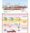
Model of aberrant G protein-coupled receptor signaling in airway smooth muscle contributing to elevated airway smooth muscle tone. Within the context of airway remodeling, G protein-coupled receptor (GPCR) signaling leading to airway smooth muscle (ASM) contraction may be altered at 3 different levels in the asthmatic airway. First, ASM may be exposed to greater levels of GPCR agonists that promote contraction (i.e., those activating Gq- or Gi-coupled receptors), or to lower levels of GPCR agonists that mediate prorelaxant signaling (Gs-coupled receptors). Increased levels of procontractile agonists can augment contraction, whereas combined stimulation of ASM with multiple Gq/Gi -coupled receptor agonists has a synergistic effect on contraction [277]. The sources of both procontractile and prorelaxant agonists include infiltrating inflammatory cells (e.g., mast cells releasing histamine, platelets releasing thrombin), postganglionic neurons with exaggerated cholinergic discharge resulting from stimulated reflex arcs or dysregulated m2 muscarinic acetylcholine receptor (mAChR) -mediated feedback inhibition [281], and resident airway cells such as epithelium, fibroblasts, and ASM itself. Access of these agonists to ASM may be increased by sloughing of airway epithelium. Second, Gq- or Gi-coupled receptor signaling may be sensitized, or Gs-coupled receptor signaling desensitized, resulting in an imbalance of signaling promoting increased phosphoinositide generation and increased calcium mobilization. Third, the contractile response to calcium may be exaggerated due to "augmented sensitization" manifested in increased myosin light chain phosphorylation caused by: 1) an imbalance of GPCR-derived signals; and 2) increased expression and activity of myosin light chain kinase (MLCK) associated with inhibited myosin light chain phosphatase (MLCP) activity, the latter a result of increased RhoA activity mediated by upregulated G12/13 and RhoA expression. Not included in this model (for the sake of simplicity) are other potential regulatory features of ASM contraction including ASM strain and load, direct contribution of non-GPCR signaling pathways to phospholipase C (PLC) activation and calcium mobilization, calcium loading in intracellular stores, compartmentalization of calcium and calcium signaling, and ion channel and membrane pump activity that directly and indirectly affect intracellular calcium levels.

References

    1. Penn RB, Pronin AP, Benovic JL. Regulation of G protein-coupled receptor kinases. Trends Cardiovasc Med. 2000;10:81–89. - PubMed
    1. Barnes PJ. Pharmacology of airway smooth muscle. Am J Respir Crit Care Med. 1998;158(Suppl):S123–S132. - PubMed
    1. Hall IP. Second messengers, ion channels and pharmacology of airway smooth muscle. Eur Respir J. 2000;15:1120–1127. - PubMed
    1. Douglas JS. Receptors on target cells. Receptors on airway smooth muscle. Am Rev Respir Dis. 1990;141(Suppl):S123–S126. - PubMed
    1. Tschumperlin DJ, Drazen JM. Mechanical stimuli to airway remodeling. Am J Respir Crit Care Med. 2001;164(Suppl):S90–S94. - PubMed

Publication types

LinkOut - more resources