. 2003 Apr;12(4):734-47.
doi: 10.1110/ps.0228503.

Noncovalent scFv multimers of tumor-targeting anti-Lewis(y) hu3S193 humanized antibody

Affiliations

Noncovalent scFv multimers of tumor-targeting anti-Lewis(y) hu3S193 humanized antibody

Barbara E Power et al. Protein Sci. 2003 Apr.

Abstract

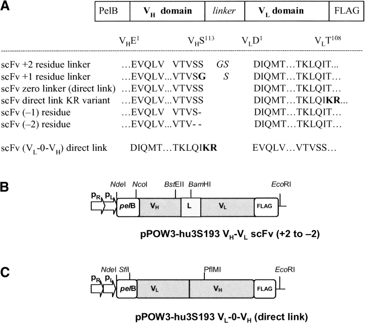
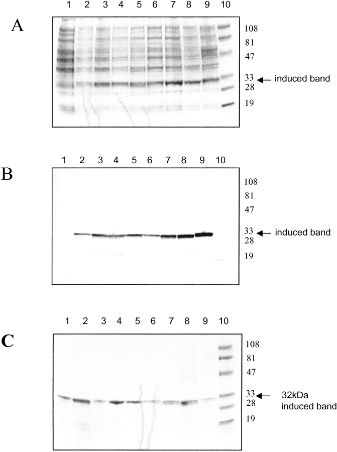
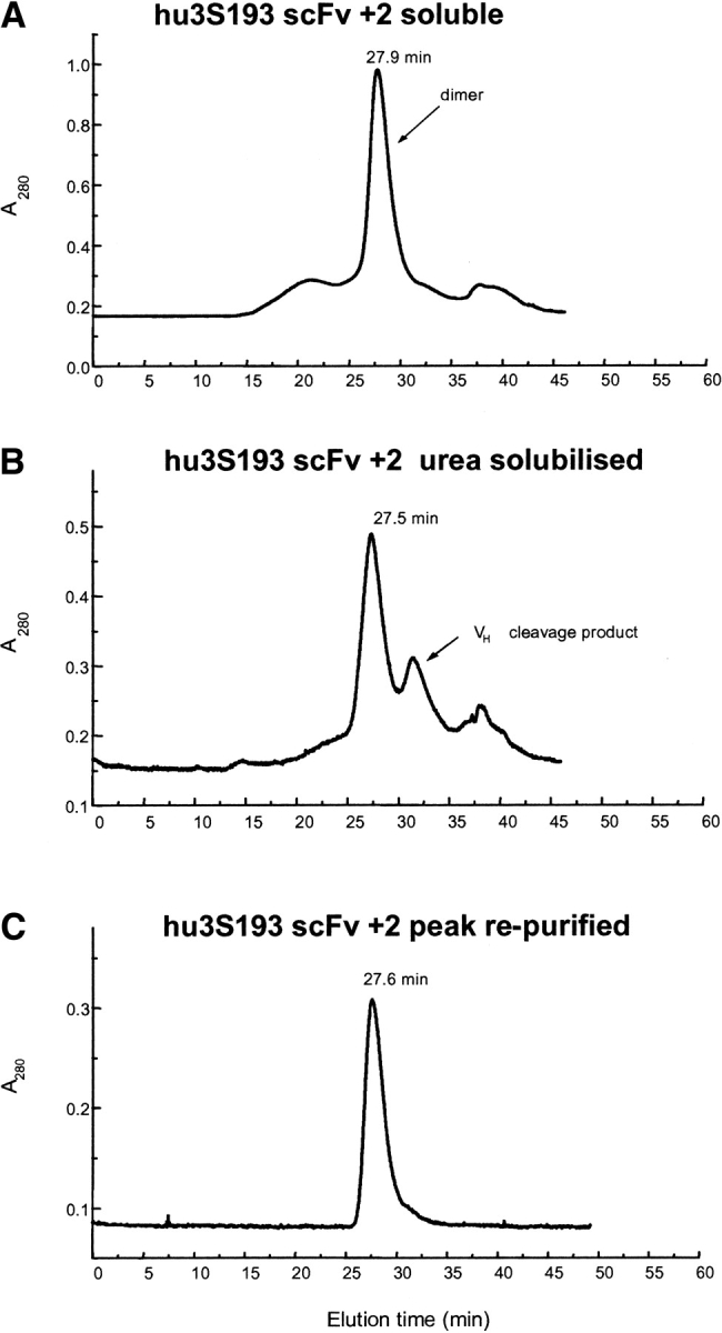
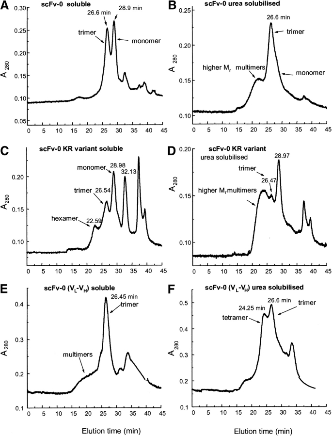
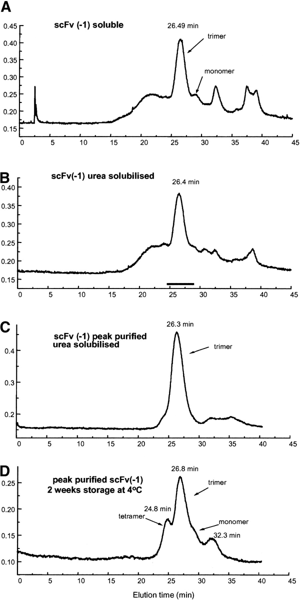
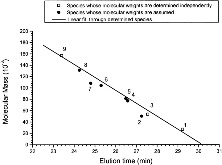
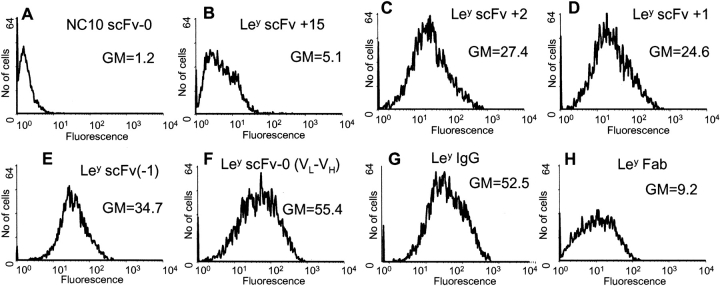
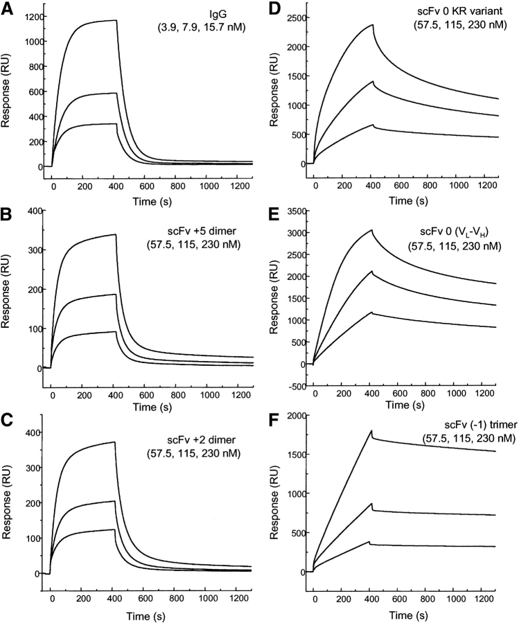
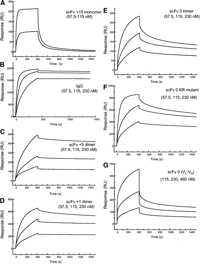
Single-chain variable fragments (scFvs) of anti-Lewis(y) hu3S193 humanized antibody were constructed by joining the V(H) and V(L) domains with either +2 residues, +1 residue, or by directly linking the domains. In addition two constructs were synthesized in which one or two C-terminal residues of the V(H) domain were removed (-1 residue, -2 residue) and then joined directly to the V(L) domain. An scFv construct in the reverse orientation with the V(L) joined directly to the V(H) domain was also synthesized. Upon transformation into Escherichia coli all scFv constructs expressed active protein. Binding activity, multimeric status, and multivalent properties were assessed by flow cytometry, size exclusion chromatography, and biosensor analysis. The results for hu3S193 scFvs are consistent with the paradigm that scFvs with a linker of +3 residues or more associate to form a non-covalent dimer, and those with a shorter linker or directly linked associate predominantly to form a non-covalent trimer and tetramer that are in equilibrium. While the association of V domains to form either a dimer or trimer/tetramer is governed by the length of the linker, the stability of the trimer/tetramer in the equilibrium mixture is dependent on the affinity of the interaction of the individual V domains to associate to form the larger Fv module.


Figures

Figure 1.
Figure 1.
Schematic diagram of the seven different hu3S193 scFv expression units. (A) Sequences of the hu3S193 scFv linker regions, with definition of the constructs demonstrating various linker lengths joining the VH and VL domains. The amino-acid sequence of the VH domain C terminus, N terminus of the VL domain and the linker residues used in each construct including the reverse orientation (VL-VH) construct are shown. (B) VH-VL and (C) VL -VH constructs in the heat-inducible pPOW3 vector, showing unique restriction sites used during the gene construction.
Figure 2.
Figure 2.
Reduced SDS-PAGE (12% polyacrylamide gels) of bacterial cultures of hu3S193 scFvs expressed using the heat-inducible pPOW3 expression vector in Escherichia coli Topp10 cells. (A) Coomassie Blue R-250 stained gel; (B) Western transfer of a duplicate gel probed with anti-FLAG FLYTAG antibody; (C) affinity-purified hu3S193 scFv multimer series (Coomassie stained). Each lane was loaded with a standardized amount of cell culture for each hu3S193 scFv multimer construct. The samples were taken at 2 h postinduction, unless otherwise indicated. Gels A and B loaded: lane 1, hu3S193 +5 (preinduction control); lane 2, hu3S193 +5; lane 3, hu3S193 +2; lane 4, hu3S193 +1; lane 5, hu3S193 direct link; lane 6, hu3S193 direct link KR variant; lane 7, hu3S193 (−1); lane 8, hu3S193 (−2); lane 9, hu3S193 reverse VL-0-VH; lane 10, low-range, molecular-weight markers (kD) 102, 81, 47, 32.7, 30, 24. Gel C contained the same samples after affinity purification, with lane 1, soluble hu3S193 +5; lane 2, hu3S193 +5 urea solubilized; all other lanes loaded as for Gels A and B.
Figure 3.
Figure 3.
Size exclusion chromatography profiles of affinity-purified hu3S193 scFv +2 on a calibrated Superose 12 HR10/30 column equilibrated in PBS, pH 7.4 and run at a flow rate of 0.5 mL/min. (A) hu3S193 +2 isolated from soluble protein fraction, predominantly dimer (54 kD); (B) urea-solubilized fraction of the cell pellet, also predominantly dimer and the VL cleavage fragment; and (C) rechromatography of the hu3S193 +2 dimer peak from panel B.
Figure 4.
Figure 4.
Size exclusion chromatography profiles of affinity purified hu3S193 direct linked (scFv-0) proteins isolated from either the soluble protein fraction or the urea-solubilized fraction. (A) hu3S193 VH-0-VL soluble, (B) hu3S193 VH-0-VL urea solubilized, (C) VH-0-VL KR variant soluble, (D) VH-0-VL KR variant urea solubilized, (E) reverse VL-0-VH soluble, and (F) reverse VL-0-VH urea-solubilized fraction.
Figure 5.
Figure 5.
Size exclusion chromatography profiles of affinity purified hu3S193 scFv (−1) isolated from (A) soluble protein fraction, predominantly as trimer (Mr ~81 kD), (B) urea-solubilized fraction, (C) rechromatography of the hu3S193 (−1) trimer peak from panel B, and (D) rechromatography of the hu3S193 (−1) trimer peak from panel B after 2 wk storage at 4°C showing formation of tetramer, monomer, and the VL cleavage fragment.
Figure 6.
Figure 6.
Relationship between molecular mass and elution time on a Superose 12 column for the family of hu3S193 scFv multimers, anti-idiotype hu3S193 antibodies F2–3A and F3–27 fragments, and the hu3S193 scFv-anti-idiotype antibody complexes formed. Samples were run on calibrated Superose 12 HR10/30 column equilibrated in PBS, pH 7.4 and run at a flow rate of 0.5 mL/min. (1) hu3S193 +15 monomer (26.8 kD), (2) anti-idiotype F2–3A Fab (50 kD), (3) hu3S193 +2 dimer (53.4 kD), (4) hu3S193 +15 monomer/F2–3A Fab complex (77 kD), (5) hu3S193 (−1) trimer (81 kD), (6) hu3S193 +5 dimer/F2–3A Fab (1:1) complex (104 kD), (7) hu3S193 (−1) tetramer (108 kD, see Fig. 5D ▶), (8) hu3S193 (−1) trimer/F2–3A Fab 1:1 complex (131 kD), (9) hu3S193 IgG (157 kD). The solid line of linear fit through the independently determined data points is plotted with R-square value = 0.991.
Figure 7.
Figure 7.
Flow cytometry analysis of hu3S193 multimers, Fab and IgG binding to Ley positive cell line, MCF-7. SK-MEL28 cells were used as an antigen negative cell line (results not shown). Cells were incubated in 100 μL of growth medium containing 5 μg hu3S193 scFv or antibody. The hu3S193 scFv multimers, Fab and IgG, were labeled with Alexa Fluor 488 and fluorescence was measured directly with a 488 argon laser. Labeled NC10 scFv-0 was used as a negative control (A). The geometric mean fluorescence (GM) is shown on each panel.
Figure 8.
Figure 8.
Sensorgrams illustrating the binding hu3S193 scFv multimers to immobilized synthetic Ley tetrasaccharide-BSA complex (2100 RU) at a constant flow rate of 5 μL/min with an injection volume of 35 μL. The surface was regenerated with 10 μL of 100 mM HCl after each cycle. (A) hu3S193 IgG (3.9, 7.9, 15.7nM), (B) hu3S193 +5 dimer (57.5, 115, 230nM), (C) hu3S193 +2 dimer (57.5, 115, 230nM), (D) VH-0-VL KR variant trimer (57.5, 115, 230nM), (E) reverse VL-0-VH trimer/tetramer (57.5, 115, 230nM), (F) hu3S193 (−1) trimer (57.5, 115, 230 nM). Monomeric forms (hu3S193 +15 and Fab) not shown, and dimeric forms (IgG and hu3S193 +5 and hu3S193 +2) exhibit monovalent binding with the surface whereas hu3S193 trimer and tetramer show multivalent binding as indicated by the dissociation rates.
Figure 9.
Figure 9.
Sensorgrams illustrating the binding of hu3S193 multimers to immobilized anti-idiotype hu3S193 antibody F2–3A (2340 RU) at a constant flow rate of 5 μL/min with an injection volume of 35 μL. The surface was regenerated with 10 μL of 100mM HCl after each cycle. All samples are shown at 57.5, 115, and 230 nM concentration. (A) hu3S193 scFv +15 monomer, (B) hu3S193 IgG, (C) hu3S193 +5 dimer, (D) hu3S193 +2 dimer, (E) VH-0-VL trimer/monomer mixture, (F) VH-0-VL [KR variant] trimer, (G) reverse VL-0-VH trimer/tetramer mixture. The hu3S193 scFv dimer and trimer peaks were isolated by size exclusion chromatography prior to biosensor analysis to remove higher molecular mass species from each multimer in the analysis.

Similar articles

Cited by

References

    1. Adams, G.P. and Schier, R. 1999. Generating improved single-chain Fv molecules for tumor targeting. J. Immunol. Methods 231 249–260. - PubMed
    1. Adams, G.P., Schier, R., McCall, A.M., Crawford, R.S., Wolf, E.J., Weiner, L.M., and Marks, J.D. 1998a. Prolonged in vivo tumour retention of a human diabody targeting the extracellular domain of human HER2/neu. Br. J. Cancer 77 1405–1412. - PMC - PubMed
    1. Adams, G.P., Schier, R., Marshall, K., Wolf, E.J., McCall, A.M., Marks, J.D., and Weiner, L.M. 1998b. Increased affinity leads to improved selective tumor delivery of single-chain Fv antibodies. Cancer Res. 58 485–490. - PubMed
    1. Adams, G.P., Shaller, C.C., Chappell, L.L., Wu, C., Horak, E.M., Simmons, H.H., Litwin, S., Marks, J.D., Weiner, L.M., and Brechbiel, M.W. 2000. Delivery of the α-emitting radioisotope bismuth-213 to solid tumors via single-chain Fv and diabody molecules. Nucl. Med. Biol. 27 339–346. - PubMed
    1. Atwell, J.L., Breheney, K.A., Lawrence, L.J., McCoy, A.J., Kortt, A.A., and Hudson, P.J. 1999. scFv multimers of the anti-neuraminidase antibody NC10: Length of the linker between VH and VL domains dictates precisely the transition between diabodies and triabodies. Protein Eng. 12 597–604. - PubMed

LinkOut - more resources