. 2003 Apr;72(4):984-97.
doi: 10.1086/374721. Epub 2003 Mar 14.

Missense mutations in the homeodomain of HOXD13 are associated with brachydactyly types D and E

Affiliations

Missense mutations in the homeodomain of HOXD13 are associated with brachydactyly types D and E

David Johnson et al. Am J Hum Genet. 2003 Apr.

Abstract

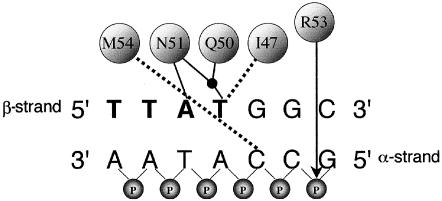
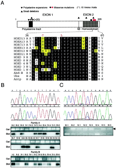
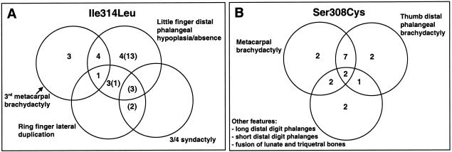
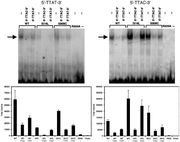
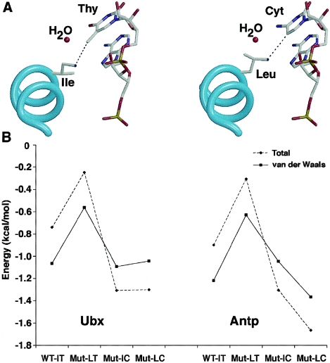
HOXD13, the most 5' gene of the HOXD cluster, encodes a homeodomain transcription factor with important functions in limb patterning and growth. Heterozygous mutations of human HOXD13, encoding polyalanine expansions or frameshifts, are believed to act by dominant negative or haploinsufficiency mechanisms and are predominantly associated with synpolydactyly phenotypes. Here, we describe two mutations of HOXD13 (923C-->G encoding Ser308Cys and 940A-->C encoding Ile314Leu) that cause missense substitutions within the homeodomain. Both are associated with distinctive limb phenotypes in which brachydactyly of specific metacarpals, metatarsals, and phalangeal bones is the most constant feature, exhibiting overlap with brachydactyly types D and E. We investigated the binding of synthetic mutant proteins to double-stranded DNA targets in vitro. No consistent differences were found for the Ser308Cys mutation compared with the wild type, but the Ile314Leu mutation (which resides at the 47th position of the homeodomain) exhibited increased affinity for a target containing the core recognition sequence 5'-TTAC-3' but decreased affinity for a 5'-TTAT-3' target. Molecular modeling of the Ile314Leu mutation indicates that this mixed gain and loss of affinity may be accounted for by the relative positions of methyl groups in the amino acid side chain and target base.


Figures

Figure  1
Figure 1
Simplified representation of binding interactions between D. melanogaster Ubx protein and a double-stranded DNA target (Passner et al. 1999). At the top, I47, Q50, N51, and M54 are the four amino acids—Ile, Gln, Asn, and Met, respectively (numbered according to their position in the homeodomain)—that contact specific bases, either through hydrogen bonds (solid lines) or hydrophobic interactions (dashed lines). The black dot represents a water molecule. Designation of the α and β strands follows Billeter (1996). The 5′-TTAT-3′ motif on the β-strand of the DNA target is shown in bold. On the α-strand only, the connecting phosphate (P) groups are included to demonstrate the position (arrow) of the ionic interaction with Arg (R53). Key amino acids in human HOXD13 are identical to those in Ubx, except that M54 is replaced by V54 (Val).
Figure  2
Figure 2
Pedigrees of families A, B, and C. Squares represent males; circles represent females; patterns of manifestation (hands only) are categorized as shown in the key at the bottom. The arrows identify the probands, and asterisks indicate individuals who underwent radiographic examination.
Figure  3
Figure 3
Missense mutations in the homeodomain of HOXD13. A, above, Schematic structure of HOXD13 showing portions encoded by each exon, the position of the splice site (sp), characteristic motifs (polyalanine tract and homeodomain), and the positions of different types of mutation (red dots identify missense mutations reported here). Below, Amino acid sequence conservation in the second half of the homeodomain. The sequence of human HOXD13 is compared with its paralogues, other members of the HOXD group, and the D. melanogaster HOM-C members Abd-B, Ubx, and Antp. The two amino acid substitutions in HOXD13 described in this report are shown above the wild-type sequence in red. Levels of conservation are indicated as follows: black, fully conserved; yellow, moderately conserved; white, poorly conserved. B, Identification of 940A→C mutation in families A and B. The upper panel shows DNA sequence chromatograms from a normal individual (above) and affected individual IV-2 from family B (below). The lower panels show confirmation of the mutation by ASO blot hybridization. C, Identification of 923C→G mutation in family C. The upper panel shows DNA sequence chromatograms from a normal individual (above) and affected individual III-2 (below). The lower panel shows confirmation of the mutation by loss of a DdeI restriction site (arrowhead).
Figure  4
Figure 4
Clinical and radiographic appearance and MCPPs of limbs in individuals heterozygous for the Ile314Leu mutation in HOXD13. A, Individual III-13 from family A. Severe middle-finger metacarpal brachydactyly is present bilaterally and confirmed on the radiograph. Note also the mild clinodactyly of the ring finger. One other hand shows a similar MCPP. B, Individual IV-3 from family B (age 5 years). Absence of the distal phalanx of the little finger is confirmed on the radiograph. Two other hands show similar MCPPs. C, Left and middle, individual II-2 from family B. Note asymmetrical abnormality of the ring fingers: on the right hand there is lateral duplication of the middle phalanx, and on the left hand, there is mild clinodactyly. In addition, the hands show features illustrated in panels A and B. C, Right, The right hand of proband IV-2 in family B (age 1 year) showing lateral duplication of the middle and distal phalanges of the ring finger. D, Foot radiograph of individual III-5 from family B. Note the unilateral shortening of the left fourth metatarsal.
Figure  5
Figure 5
Combinations of individual abnormalities in hands from subjects with the Ile314Leu mutation (A) and the Ser308Cys mutation (B). In A, figures in parentheses refer to additional cases classified clinically but not radiographically.
Figure  6
Figure 6
In vitro binding of wild-type (WT) and mutant (I314L, S308C, and R320A) HOXD13 proteins to synthetic ds oligonucleotides. Above, representative gel shift assays employing 32P-labeled 5′-TTAT-3′ probe (left) and 5′-TTAC-3′ probe (right) in the absence (−) or presence of unlabeled competitor oligonucleotides. The arrow shows the position of the bound oligonucleotide/protein complexes. Below, quantitation (mean ± SEM) of total counts from 4–5 experiments. Note the difference in scale of absolute counts on the Y-axis.
Figure  7
Figure 7
Interaction between HOXD13 and DNA sequences. A, Models based on the Ubx/DNA complex, highlighting the interaction between Ile and thymine (Thy) in the wild type (left panel) and between Leu and cytosine (Cyt) in the “double mutant” (right panel). The cyan spiral indicates the main chain of homeodomain helix III; atoms (except H) in the position 47 side chain and DNA are color coded as follows: C = gray; O = red; N = dark blue; P = orange. B, Total (dashed line) and van der Waals (solid line) interaction energies (in kcal/mol) between Ile and Thy (WT-IT, wild type), Leu and Thy (Mut-LT, Ile→Leu mutant), Ile and Cyt (Mut-IC, Thy→Cyt mutant), and Leu and Cyt (Mut-LC, double mutant). Models were built using the Ubx (left) and Antp (right) homeodomain/DNA complexes.

References

Electronic-Database Information

    1. Ensembl, http://www.ensembl.org/Homo_sapiens/mapview?chr=2 (for physical map of 2q31 region)
    1. GenBank, http://www.ncbi.nlm.nih.gov/GenBank/ (for HOXD13 [accession numbers. AF005219, AF005220, AC009336, and NM_000523])
    1. Homeodomain Resource, http://research.nhgri.nih.gov/homeodomain/ (for homeodomain sequences and DNA binding sites)
    1. Online Mendelian Inheritance in Man (OMIM), http://www.ncbi.nlm.nih.gov/Omim/(for SPD, HFUS, BDD, and BDE)
    1. Protein Data Bank, http://www.pdb.org/ (for Antp [ID: 9ANT] and Ubx [ID: 1B8I] homeodomain-DNA structures)

References

    1. Albrecht AN, Schwabe GC, Stricker S, Böddrich A, Wanker EE, Mundlos S (2002) The synpolydactyly homolog (spdh) mutation in the mouse: a defect in patterning and growth of limb cartilage elements. Mech Dev 112:53–67 - PubMed
    1. Armour CM, Bulman DE, Hunter AGW (2000) Clinical and radiological assessment of a family with mild brachydactyly type A1: the usefulness of metacarpophalangeal profiles. J Med Genet 37:292–296 - PMC - PubMed
    1. Banerjee-Basu S, Baxevanis AD (2001) Molecular evolution of the homeodomain family of transcription factors. Nucleic Acids Res 29:3258–3269 - PMC - PubMed
    1. Banerjee-Basu S, Sink DW, Baxevanis AD (2001) The Homeodomain Resource: sequences, structures, DNA binding sites and genomic information. Nucleic Acids Res 29:291–293 - PMC - PubMed
    1. Bell J (1951) On brachydactyly and symphalangism. In: The treasury of human inheritance. Vol 5. Cambridge University Press, Cambridge, pp 1–31

Publication types

MeSH terms

Associated data