. 2003 Apr;162(4):1163-74.
doi: 10.1016/S0002-9440(10)63912-0.

Role of CD31/platelet endothelial cell adhesion molecule-1 expression in in vitro and in vivo growth and differentiation of human breast cancer cells

Affiliations

Role of CD31/platelet endothelial cell adhesion molecule-1 expression in in vitro and in vivo growth and differentiation of human breast cancer cells

Luisella Righi et al. Am J Pathol. 2003 Apr.

Abstract

Breast ductal carcinoma in situ is an intraductal proliferation of malignant epithelial cells that diffuse within the ductal system without stromal invasion. Our finding that a subset of these tumors express CD31/platelet endothelial cell adhesion molecule-1 suggests that breast cancer represents an informative model for studying the involvement of the molecule in the morphogenesis, differentiation, and diffusion of this disease. Transfection of CD31 in MDA-MB-231 cells caused reduction in growth, loss of CD44, and acquisition of a ductal morphology. The same effects were maintained in vivo, in which CD31(+) tumors grew with in situ-like aspects, papillary differentiation, and a secretory phenotype. CD44 was down-modulated, with the CD31(+) cells blocked in the G(1) phase. The morphology was highly similar to what was observed in some human CD31(+) ductal carcinomas in situ. MDA-MB-231 mock cells grew in solid sheets, lacking stromal material, and displaying high levels of CD44 and proliferation. CD31(+) cells acquired motility characteristics in in vitro assays, a finding confirmed in vivo by the diffusion of human tumor cells throughout the normal ducts residual in the murine mammary gland. In conclusion, CD31 expression reverts the undifferentiated morphology and aggressive behavior of MDA-MB-231 cells, indicating its active role in the morphogenesis of breast ductal in situ carcinomas.


Figures
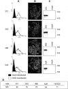
Figure 1.
Figure 1.
Preparation of CD31+ clones of the MDA-MB-231 cell line. Stable transfection by electroporation of a plasmid containing the full-length human CD31 gene results in the production of three MDA-MB-231 clones (2C1, 3D2, and 4B5) and a bulk preparation of cells homogeneously expressing CD31, as determined by examining cells in suspension (A, empty profiles) and in adherence (B). Western blot analysis highlights a single chain of ∼130 kd in the MDA-MB-231 cells transfected with CD31 (C). Control MDA-MB-231 mock cells do not show detectable levels of CD31 (A, full panels, and C, mock lanes). A: Cells were analyzed using a FACSort equipment and scoring 10,000 events/sample. x axis, fluorescence intensity/cells; y axis, number of cells registered/channel. B: Cells were grown on glass coverslips, fixed using methanol and acetone, and stained. C: Cells were lysed using 1% Nonidet P-40, run on 10% sodium dodecyl sulfate-polyacrylamide gel electrophoresis, transblotted onto a nitrocellulose membrane, and the reactivity was examined using JC70 mAb and horseradish peroxidase-conjugated GαMIg. Molecular weight markers are indicated on the right. Original magnifications, ×60.
Figure 2.
Figure 2.
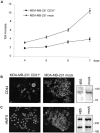
In vitro effects of CD31 transfection. CD31 transfection reduces MDA-MB-231 growth rate (A). MDA-MB-231 CD31+ 2C1, 3D2, and 4B5 clones and mock cells were seeded in complete medium, detached, and counted after 4 to 7 days by three independent observers. Fold increase was calculated as the ratio between the mean cell numbers scored and the number of cells plated at the beginning of the experiments. Data are the mean ± SD (vertical bars) from four independent experiments. CD31 transfection strongly down-modulates CD44 expression (B, clone 4B5), as examined by immunofluorescence and Western blot analysis. Moreover, it induces cytoplasmic organization of HMFG protein in secretory vacuoles (C, clone 4B5). No significant differences were seen in terms of protein quantity (C). Cells were grown on glass coverslips, fixed, stained, and photographed. Representative images from five independent experiments obtained using three different clones as well as a bulk population of MDA-MB-231 CD31+ cells. For Western blot analysis, cells were lysed using 1% Nonidet P-40, run on a 10% (CD44) or 7.5% (HMFG) sodium dodecyl sulfate-polyacrylamide gel electrophoresis, transblotted onto a nitrocellulose membrane, and the reactivity examined using JC70 mAb and horseradish peroxidase-conjugated GαMIg. Molecular weight markers are indicated on the right. Original magnification, ×60 (C).
Figure 3.
Figure 3.
MDA-MB-231 CD31+ cells form ductal and papillary-like structures in tri-dimensional cultures. Cells were seeded in a collagen gel matrix and observed daily by phase-contrast microscopy. After 2 weeks the gels were fixed and stained by H&E. By day 4 ∼80% of MDA-MB-231 CD31+ cells acquire ductal-like structures (A, clone 2C1), with occasional papillae growing inside the ducts (B, clone 2C1). Mock cells grow in a disorganized way and form solid aggregates (C and D). Original magnifications, ×100.
Figure 4.
Figure 4.
CD31 expression decreases growth in vivo. MDA-MB-231 CD31+ and mock cells were injected in the mammary foot pads of female SCID mice (arrows) and pictures were taken after 6 weeks (A). The size of the lesions at different time intervals is reported in mm in the table, and the different clones are shown (B). Data are the mean from two animals for each clone and for the bulk preparation of CD31+ cells, while they represent the mean ± SD from four animals in the MDA-MB-231 mock cells.
Figure 5.
Figure 5.
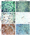
In vivo effects of CD31 expression. Histology of tumor samples derived from animals injected with MDA-MB-231 CD31+ or mock cells. Trichrome stain shows that MDA-MB-231 CD31+ tumors are made of cells organized in ductal structures and surrounded by thick hyaline stroma. The ducts are partly occupied by tumor cells forming papillae; blood vessels are evident (A). Immunohistochemistry indicates that the basal membrane of CD31+ tumors is made of collagen IV (C) and laminin (E). The lesions derived from mock cells grow in solid laminae (B), with minimal amounts of stroma (D and F). The figure shows the histology of a tumor derived from clone 4B5. Original magnifications, ×40.
Figure 6.
Figure 6.
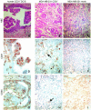
The tumors obtained from MDA-MB-231 CD31+ cells grown in SCID mice were compared to a human sample selected as paradigmatic of a population of CD31+ breast cancers. H&E staining performed after 6 weeks of growth in SCID mice shows that the MDA-MB-231 CD31+ tumors preferentially expand within the ducts, forming papillae (B), similar to what observed in human CD31+ DCIS (A). Conversely, MDA-MB-231 mock tumors constantly grow in solid lamina (C). The immunophenotype of CD31+ SCID mice samples matches the one observed in human CD31+ DCIS. The CD31 molecule is expressed at the cell membrane level, with the highest degree of positivity scored by the cells forming papillae (D, human lesions; E, murine lesions). The basal cells lining the lumen focally express CD44 (H, arrow), in analogy to human CD31+ DCIS (G). MDA-MB-231 mock tumors maintained the phenotype of the original line, ie, CD31 and CD44+, which was unaltered during the whole observation (F and I). The pictures were obtained from a selection of lesions derived from mice injected with MDA-MB-231 CD31+ cells (clones 2C1 and 4B5). Original magnifications, ×40.
Figure 7.
Figure 7.
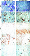
CD31 expression induces differentiation of MDA-MB-231 cells in vivo. PAS staining highlights intracytoplasmic vacuoles only in tumors derived from transfected cells and mainly in the cells forming the papillae (A, arrows; and B). MDA-MB-231 CD31+ tumors acquire a secretory phenotype, as inferred by the presence of HMFG protein in cytoplasmic vacuoles (large arrows) and in some ductal lumens (small arrows) (C). Mock cells display scattered intracytoplasmic HMFG protein granules (arrows), without evidence of secretion (D). MDA-MB-231 CD31+ tumor cells are not actively proliferating. Ki-67 index is <1% in MDA-MB-231 CD31+ tumors (E), whereas p27 is highly expressed (>85%) (G), as are normal murine residual glands (large arrows in G and H) and normal stromal murine cells (small arrows in G and H). Conversely, mock tumors score >90% for Ki-67 (F) and <50% for p27 (H). Nuclear p21 was seen in <1% of the MDA-MB-231 CD31+ cells and was undetectable within the cytoplasm, implying that the cells are blocked in the G1 phase (I). MDA-MB-231 mock tumors express nuclear p21 in ∼10% of the cells (large arrows in L), whereas generalized faint cytoplasmic staining was observed in the remaining population (small arrows). The pictures were obtained from a selection of lesions derived from mice injected with MDA-MB-231 CD31+ cells (clones 2C1 and 4B5). Original magnifications: ×40 (A–D); ×20 (E–L).
Figure 8.
Figure 8.
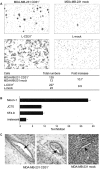
CD31 confers unique kinetic properties to MDA-MB-231 cells. The presence of CD31 is a prerequisite for MDA-MB-231 cells to migrate in in vitro assays (A, top) as well as for murine L-fibroblasts (A, bottom). The table shows cumulative data from several experiments. The asterisks indicate statistical significance (P < 0.05, Wilcoxon matched-pairs signed-ranks test). Migration is blocked only by the Moon-1 mAb (reactive to the CD31 domain 2), whereas other mAbs (reactive to the CD31 domain 1) are only slightly effective. Vertical bars represent the mean ± SD from four independent experiments (B). The unique migratory properties of MDA-MB-231 CD31+ cells are characterized in vivo by the tendency to invade the normal murine residual mammary ducts maintaining the original architecture, as compared to the compression and destruction of the same structures evident in the lesions originated from mock cells (C, arrows; clone 4B5).

Similar articles

Cited by

References

    1. DeLisser HM, Newman PJ, Albelda SM: Molecular and functional aspects of PECAM-1/CD31. Immunol Today 1994, 15:490-495 - PubMed
    1. Muller WA, Weigl SA, Deng X, Phillips DM: PECAM-1 is required for transendothelial migration of leukocytes. J Exp Med 1993, 178:449-460 - PMC - PubMed
    1. Muller WA, Randolph GJ: Migration of leukocytes across endothelium and beyond: molecules involved in the transmigration and fate of monocytes. J Leukoc Biol 1999, 66:698-704 - PubMed
    1. Lu TT, Yan LG, Madri JA: Integrin engagement mediates tyrosine dephosphorylation on platelet-endothelial cell adhesion molecule 1. Proc Natl Acad Sci USA 1996, 93:11808-11813 - PMC - PubMed
    1. Piali L, Hammel P, Uherek C, Bachmann F, Gisler RH, Dunon D, Imhof BA: CD31/PECAM-1 is a ligand for alpha v beta 3 integrin involved in adhesion of leukocytes to endothelium. J Cell Biol 1995, 130:451-460 - PMC - PubMed

Publication types

Substances