. 2003 Apr;162(4):1221-7.
doi: 10.1016/S0002-9440(10)63918-1.

Expression and regulation of Toll-like receptor 2 in rheumatoid arthritis synovium

Affiliations

Expression and regulation of Toll-like receptor 2 in rheumatoid arthritis synovium

Reinhart Seibl et al. Am J Pathol. 2003 Apr.

Abstract

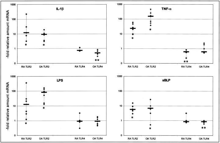
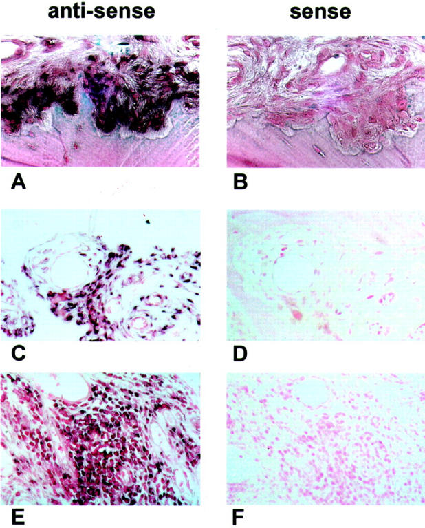
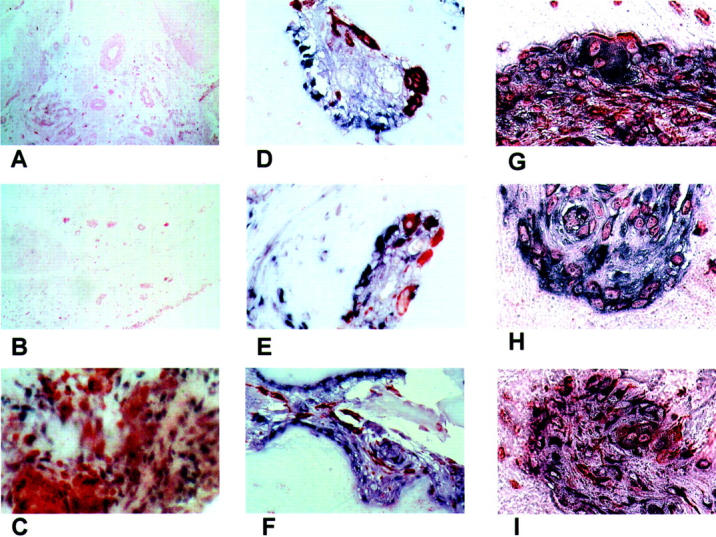
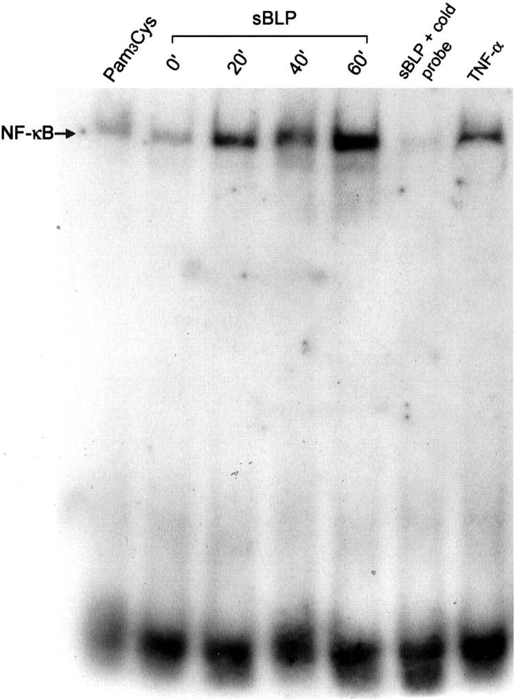
Toll-like receptors (TLRs) are involved in mediating cell activation on stimulation with microbial constituents. We investigated the role for TLRs in synovial fibroblast (SF) activation in rheumatoid arthritis (RA). We analyzed whether stimulation with interleukin-1 beta and tumor necrosis factor-alpha, cytokines present in RA synovium, influences expression of TLR genes in SFs. The effects were compared with those of treatment with lipopolysaccharide and a synthetic lipopeptide (sBLP). Gene expression was examined using quantitative polymerase chain reaction. TLR2-mediated cell activation was investigated by electromobility shift assay for nuclear factor-kappa B. To localize TLR2 expression in joint tissue sections of RA patients were stained using in situ hybridization. Expression of TLR2 in RA SFs was increased after treatment with interleukin-1 beta, tumor necrosis factor-alpha, lipopolysaccharide, and sBLP. Nuclear factor-kappa B translocation in SFs was triggered by TLR2-mediated cell stimulation. Synovial tissues from RA joints expressed TLR2 predominantly at sites of attachment and invasion into cartilage and bone. The observed elevated expression of TLR2 in RA SFs could be a consequence of direct exposure to microbial compounds or of the presence of inflammatory mediators in the joint. TLR-associated signaling pathways may contribute to the pathogenesis of RA, either by initiating or perpetuating activation of SFs.


Figures

Figure 1.
Figure 1.
Effects of stimulation on the expression of TLR2 and TLR4 mRNA in RA (n = 6) and OA (n = 7) SFs. The ordinate shows the relative increase of specific mRNA compared to nonstimulated cells. Each symbol indicates one cell culture from one RA patient examined. The bars indicate the medians. The increase of TLR2 mRNA in response to all four stimuli reached statistical significance (*, Wilcoxon paired-sample test; tied Z values from −2.37 to −2.02, P values from 0.02 to 0.04). Levels of TLR4 mRNA expression of stimulated cells did not differ significantly from nonstimulated cells apart from stimulation of RA SFs with TNF-α, and from stimulation of OA SFs with IL-1β or sBLP (**, Wilcoxon paired-sample test; tied Z values from−2.2 to −2.36, P values from 0.02 to 0.04).
Figure 2.
Figure 2.
TLR2 in situ hybridizations of RA synovial tissues. One representative section of RA synovial tissue of seven patients, hybridized in situ with specific RNA probes for TLR2 mRNA (A, C, E). Cells positive for TLR2 mRNA are dark purple. As negative controls, corresponding tissue sections were hybridized with the sense probes. All tissues hybridized with the sense control probes show no specific signals (B, D, F). Cells expressing detectable levels of TLR2 mRNA are located in areas of adhesion and invasion into bone or cartilage (A; negative control, B), around small vessels (C; negative control, D) and in areas of lymphocyte infiltrations (E; negative control, F). Original magnifications: ×200 (A, B, C, E); ×100 (D, F).
Figure 3.
Figure 3.
Control and double stainings. A: One representative section from OA tissues of four patients stained for TLR2 mRNA. B: One representative tissue section from one normal subject of two stained for TLR2 mRNA. Representative tissue section derived from RA-synovium, stained with anti-CD3 antibodies (specific for T cells) after in situ hybridization with a probe specific for TLR2 mRNA. TLR2 mRNA presents as purple/brown color, T cells are red. C: Expression of TLR2 mRNA and CD3 is detectable in distinct cells. Representative tissue sections derived from RA-synovium at sites of synovial invasion into cartilage. The sections are stained with anti-CD68 antibodies (specific for macrophages) after in situ hybridization with probes specific for TLR2 mRNA. TLR2 mRNA presents as purple color, macrophages are red. D–F: Most of the cells expressing TLR2 mRNA are not expressing CD68. Representative tissue section derived from RA synovium at sites of synovial invasion into cartilage. The sections are stained with antibodies against vimentin (G–H) or prolyl-4-hydroxylase (I) after in situ hybridization with probes specific for TLR2 mRNA. TLR2 presents as purple/black color, antibodies are visible as orange. A great portion of cells expressing TLR2 is positive for the fibroblast markers. Original magnifications: ×100 (A, B); ×200 (C); ×630 (D, E, G, I); ×400 (F).
Figure 4.
Figure 4.
Nuclear translocation of NF-κB after TLR2-dependent stimulation. Representative EMSA for examining the effects of stimulation with sBLP on the nuclear translocation of NF-κB in RA SFs. Three different time points (20 minutes, 40 minutes, 60 minutes) after stimulation are shown compared to nonstimulated cells (0 minutes), cells exposed to Pam3Cys, or stimulated with TNF-α. The specificity of NF-κB binding to the oligonucleotides was confirmed by the addition of unlabeled probe (cold probe) to the binding reaction. The data show one representative experiment of three.

References

    1. Rock FL, Hardiman G, Timans JC, Kastelein RA, Bazan JF: A family of human receptors structurally related to Drosophila Toll. Proc Natl Acad Sci USA 1998, 95:588-593 - PMC - PubMed
    1. Takeuchi O, Kawai T, Sanjo H, Copeland NG, Gilbert DJ, Jenkins NA, Takeda K, Akira S: TLR6: a novel member of an expanding Toll-like receptor family. Gene 1999, 231:59-65 - PubMed
    1. Du X, Poltorak A, Wei Y, Beutler B: Three novel mammalian Toll-like receptors: gene structure, expression, and evolution. Eur Cytokine Netw 2000, 11:362-371 - PubMed
    1. Aderem A, Ulevitch RJ: Toll-like receptors in the induction of the innate immune response. Nature 2000, 406:782-787 - PubMed
    1. Muzio M, Bosisio D, Polentarutti N, D’Amico G, Stoppacciaro A, Mancinelli R, van’t Veer C, Penton-Rol G, Ruco LP, Allavena P, Mantovani A: Differential expression and regulation of Toll-like receptors (TLR) in human leukocytes: selective expression of TLR3 in dendritic cells. J Immunol 2000, 164:5998-6004 - PubMed

Publication types

MeSH terms

LinkOut - more resources