Comparative Study
. 2003 Apr;162(4):1361-71.
doi: 10.1016/S0002-9440(10)63932-6.

The relationship between hypomethylation and CpG island methylation in colorectal neoplasia

Affiliations
Comparative Study

The relationship between hypomethylation and CpG island methylation in colorectal neoplasia

Carolyn Bariol et al. Am J Pathol. 2003 Apr.

Abstract

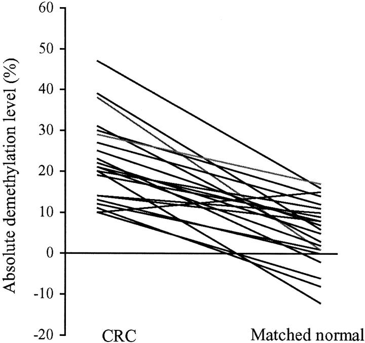
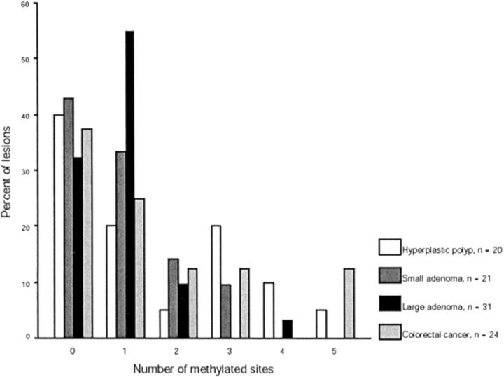
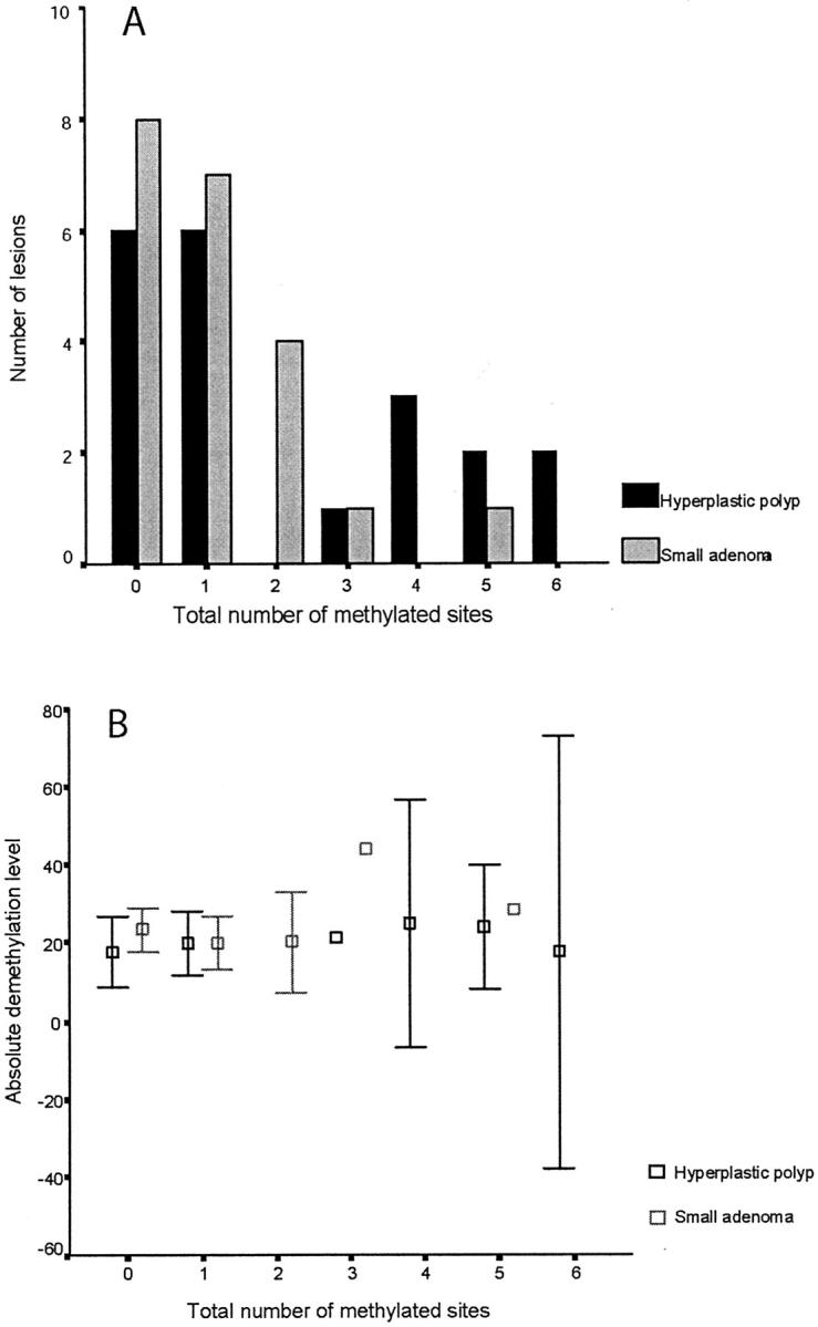
Tumors are often characterized by an imbalance in cytosine methylation as manifested both by hypermethylation of CpG islands and by genome hypomethylation. These epigenetic changes were assessed in colorectal neoplasia to determine whether they arose through a common mechanism or indeed were distinct and unrelated phenomena. Fresh representative samples of adenomas, hyperplastic polyps, colorectal cancers, and normal mucosa were used in this study. Global methylation levels were measured by analyzing the methyl-accepting capacity of DNA. Methylation of p16, hMLH1, and MINT 1, 2, 12, and 31 were assessed by bisulfite polymerase chain reaction. Microsatellite status was determined by polymerase chain reaction using six markers and hMLH1 and proliferating cell nuclear antigen expression was assessed by immunohistochemistry. Normal colonic mucosa had a higher endogenous 5-methyl cytosine content than all proliferative lesions of the colon (P < 0.001). The extent of demethylation in hyperplastic polyps and adenomas was significantly related to its proliferative rate. Right-sided hyperplastic polyps were more likely to be methylated than adenomas (odds ratio, 2.3; confidence interval, 1.1 to 4.6). There was no relationship between the level of global hypomethylation and hypermethylation. Some hyperplastic colorectal polyps have a propensity to develop dense CpG island methylation. Hypermethylation and hypomethylation contribute separately to the process of carcinogenesis.


Figures

Figure 1.
Figure 1.
Comparison of the extent of global DNA hypomethylation in the different tissue types assessed in this study. Mucosa-H, normal colonic mucosa from healthy individuals; mucosa-C, normal mucosa from individuals with neoplastic lesions; HP, hyperplastic polyps; small adenomas, adenomas ≤10-mm maximum diameter; large adenomas, adenomas >10-mm maximum diameter; colorectal cancer, colorectal carcinoma; n, number of samples analyzed in each group. For the purpose of analysis, normal tissues from patients with adenomas (n = 3) were grouped with those from patients with colorectal cancers (n = 22). The absolute demethylation level refers to the percentage of methylated CpG in the colonic sample after subtraction from the control peripheral blood mononuclear cells sample. Each sample was assayed in triplicate and the mean decrease in absolute methylation content and 95% confidence intervals are shown.
Figure 2.
Figure 2.
Comparison of methylation levels in 22 colorectal carcinomas and matched normal colonic mucosa of individual patients. Each line demonstrates the relationship between the methylation levels in the colorectal tumor and its paired normal mucosa.
Figure 3.
Figure 3.
Representative gel showing the analysis of four tumors (T1 to T3) for methylation at p16 (A) and hMLH1 regions A and C (B). For p16, bisulfite-modified DNA was amplified in separate reactions to identify unmethylated (U) and methylated template (M). All amplicons generated by the methylation-specific p16 primers were digested with BstUI. The methylated amplicon from T2 (UD) has digested (D), confirming that this tumor was truly methylated at p16. There was no amplification with the methylation-specific primers for tumor T1 and therefore this tumor is unmethylated. Methylation of hMLH1 regions A and C (B) was detected by PCR amplification of bisulfite-modified DNA followed by digestion with BstUI, which only cuts the methylated amplicon. Undigested amplicon is shown (UD), and after digestion it is apparent that T1 is unmethylated at both regions, T3 at region C only and T2 is methylated at both regions A and C. Molecular weight marker (MW) is pUC19/MspI.
Figure 4.
Figure 4.
Distribution of CpG island methylation in hyperplastic polyps, adenomas, and colorectal carcinomas. The histogram shows the percentage of methylated loci according to the histological type of the lesion. The MINT sites and p16 are included in the analysis. It is apparent that both small and large adenomas show a skewed distribution of methylation with 98% (51 of 52) of lesions demonstrating methylation at less than four sites. Hyperplastic polyps and colorectal cancer demonstrate a more continuous distribution of methylation with 6 of 44 lesions (14%) showing methylation at more than three sites.
Figure 5.
Figure 5.
Comparison of CpG island methylation (A) and corresponding levels of hypomethylation (B) in hyperplastic polyps (n = 20) and small adenomas (n = 21). The histogram shows the number of methylated loci according to the histological type of the lesion. The MINT sites, p16, hMLH1-A, and hMLH1-C are included in the analysis, so the maximum total number of methylated sites for each lesion is seven. The absolute demethylation level refers to the percentage of methylated CpG in the sample after subtraction from the control peripheral blood mononuclear cell sample. Each sample was assayed in triplicate, and the mean decrease in absolute methylation content and 95% confidence intervals are shown. There was no relationship between the number of methylated sites and the extent of demethylation for hyperplastic polyps (Spearman’s rho, 0.32; P = 0.2) or small adenomas (Spearman’s rho, 0.08; P = 0.74).

Comment in

References

    1. Ehrlich M: DNA methylation in cancer: too much, but also too little. Oncogene 2002, 21:5400-5413 - PubMed
    1. Gama-Sosa MA, Slagel VA, Trewyn RW, Oxenhandler R, Kuo KC, Gehrke CW, Ehrlich M: The 5-methylcytosine content of DNA from human tumors. Nucleic Acids Res 1983, 11:6883-6894 - PMC - PubMed
    1. Feinberg AP, Gehrke CW, Kuo KC, Ehrlich M: Reduced genomic 5-methylcytosine content in human colonic neoplasia. Cancer Res 1988, 48:1159-1161 - PubMed
    1. Toyota M, Ahuja N, Ohe-Toyota M, Herman JG, Baylin SB, Issa JP: CpG island methylator phenotype in colorectal cancer. Proc Natl Acad Sci USA 1999, 96:8681-8686 - PMC - PubMed
    1. Sharrard RM, Royds JA, Rogers S, Shorthouse AJ: Patterns of methylation of the c-myc gene in human colorectal cancer progression. Br J Cancer 1992, 65:667-672 - PMC - PubMed

Publication types

MeSH terms