. 2003 Mar 15;17(6):711-6.
doi: 10.1101/gad.1065603.

Human and mouse TPIT gene mutations cause early onset pituitary ACTH deficiency

Affiliations

Human and mouse TPIT gene mutations cause early onset pituitary ACTH deficiency

Anne-Marie Pulichino et al. Genes Dev. .

Abstract

Tpit is a highly cell-restricted transcription factor that is required for expression of the pro-opiomelanocortin (POMC) gene and for terminal differentiation of the pituitary corticotroph lineage. Its exclusive expression in pituitary POMC-expressing cells has suggested that its mutation may cause isolated deficiency of pituitary adrenocorticotropin (ACTH). We now show that Tpit-deficient mice constitute a model of isolated ACTH deficiency (IAD) that is very similar to human IAD patients carrying TPIT gene mutations. Through genetic analysis of a panel of IAD patients, we show that TPIT gene mutations are associated at high frequency with early onset IAD, but not with juvenile forms of this deficiency. We identified seven different TPIT mutations, including nonsense, missense, point deletion, and a genomic deletion. This work defines congenital early onset IAD as a relatively homogeneous clinical entity caused by recessive transmission of loss-of-function mutations in the TPIT gene.


Figures
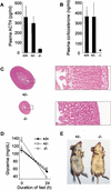
Figure 1
Figure 1
Tpit−/− mice are a model of isolated ACTH deficiency (IAD). (A) Plasma ACTH in Tpit−/− mice (n = 7) is greatly reduced compared with wild-type (+/+, n = 6) and heterozygous (+/−, n = 6) mice. (B) Plasma corticosterone is undetectable in Tpit−/− (n = 11) mice, but normal levels are observed in wild-type (n = 8) and heterozygous (n = 10) mice. (C) Tissue sections stained with hematoxylin and eosin showing hypoplastic Tpit−/− adrenals compared with wild type. (D) Fasting-induced hypoglycemia is greater in Tpit−/− (n = 12) than in wild-type (n = 9) or heterozygous (n = 12) mice. Basal glycemia was significantly higher (p ≤ 0.05) in Tpit−/− mice. After a 24-h fast, the glycemia of Tpit−/− mice is significantly lower than for other mice (p ≤ 0.001). Data are means ± S.E.M. (E) Ventral views of +/− and −/− Tpit mice. The mutant has a yellow pigmentation compared with the light gray of +/− or +/+ mice.
Figure 2
Figure 2
Human TPIT gene mutations in early onset isolated ACTH deficiency (IAD). (A) Summary of different TPIT mutations found in cases of early onset IAD. (B) DNA sequence and pedigree of patients with TPIT mutations. For each family, the black symbol represents the mutant allele indicated at top and revealed by DNA sequencing at bottom. Double horizontal bars in pedigree represent consanguinity. The gray allele in family V has a deletion of exons 3 and 4 as shown in C, and the patient is a compound heterozygote. This patient and her sibling were described previously (Malpuech et al. 1988; Dechelotte et al. 1994). (C) Southern blot analysis of genomic DNA from family V (patient and mother) showing a deletion of exons 3 and 4 in one allele of both subjects. (Lanes 1,2) Control (lane 1) and patient (lane 2) DNA digested with HindIII and hybridized with a probe for exons 3, 4, 5, and 6. A new band of 4.6 kb can be observed (lane 2). (Lanes 3,4,5,6) DNA digested with BamHI and hybridized with an exon 2 probe. In lanes 4 and 6 (patient and mother, respectively), a new band of 3.5 kb is observed compared with controls (lanes 3,5). Schematic representation indicates position of the 5.2-kb deletion in the mutant allele.
Figure 3
Figure 3
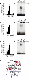
TPIT missense mutations cause loss-of-function for DNA binding. The activity (A,C,E) and DNA-binding ability (B,D,F) of the three missense mutations identified in TPIT patients was assessed. The S128F (C) and I171T (E) mutations are devoid of transcriptional activity, whereas the T58A (A) mutation retains ∼10% transcriptional activity when assayed by transient transfection into GH3 cells using a Tpit/Pitx-dependent reporter plasmid. Data are means ± S.E.M. of three to five experiments, each performed in duplicate. DNA-binding ability of the three mutant proteins was assessed by gel retardation using in vitro translated proteins and a palindromic consensus binding site of T-box proteins as probe. Synthesis of equivalent amounts of mutant and wild-type proteins was assessed by Western blot (top of each panel). No binding was observed for mutants S128F (D) and I171T (F), whereas slight binding was retained for mutant T58A (B). (G) The position of TPIT residues identified in missense IAD patients were modeled onto the crystal structure of the brachyury T-box (Muller and Herrmann 1997).

Comment in

References

    1. Argente J, Perez-Jurado L, Sotos J. Molecular bases of pathological growth. J Endocr Genet. 2000;1:179–210.
    1. Bertagna X. Proopiomelanocortin-derived peptides. Endocrinol Metab Clin North Am. 1994;23:467–485. - PubMed
    1. Chowdhary BP, Gustavsson I, Wikberg JE, Chhajlani V. Localization of the human melanocortin-5 receptor gene (MC5R) to chromosome band 18p11.2 by fluorescence in situ hybridization. Cyto Genet Cell Genet. 1995;68:79–81. - PubMed
    1. Cohen LE, Radovick S. Molecular basis of combined pituitary hormone deficiencies. Endocr Rev. 2002;23:431–442. - PubMed
    1. Dattani MT, Martinez-Barbera JP, Thomas PQ, Brickman JM, Gupta R, Martensson IL, Toresson H, Fox M, Wales JK, Hindmarsh PC, et al. Mutations in the homeobox gene HESX1/Hesx1 associated with septo-optic dysplasia in human and mouse. Nat Genet. 1998;19:125–133. - PubMed

Publication types

MeSH terms