. 2003 Mar 15;17(6):738-47.
doi: 10.1101/gad.1065703.

Tpit determines alternate fates during pituitary cell differentiation

Affiliations

Tpit determines alternate fates during pituitary cell differentiation

Anne-Marie Pulichino et al. Genes Dev. .

Abstract

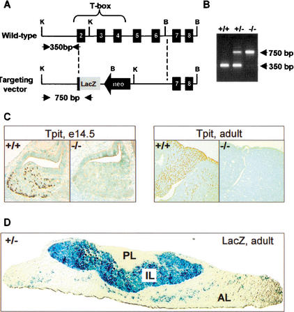
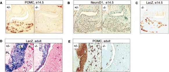
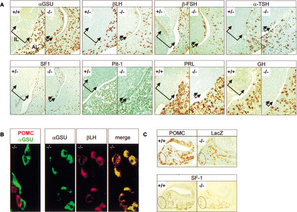
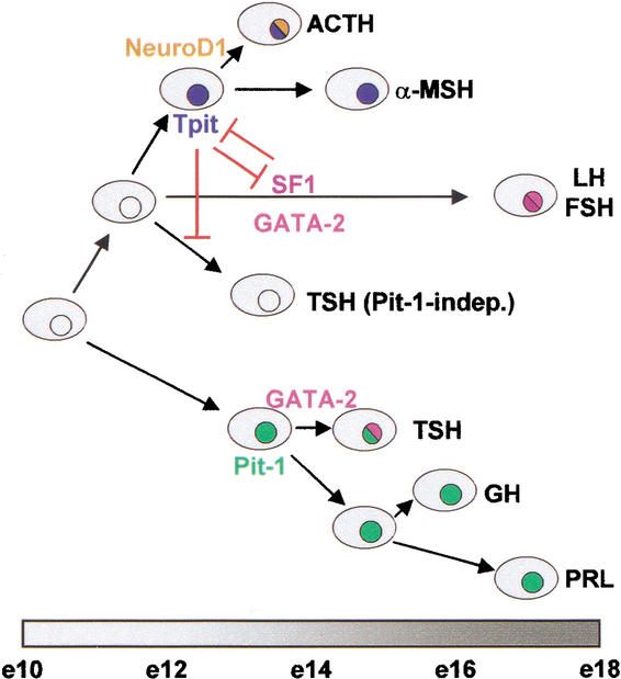
The T-box transcription factor Tpit was identified as a cell-specific factor for expression of the pituitary proopiomelanocortin (POMC) gene. Expression of this factor is exclusively restricted to the pituitary POMC-expressing lineages, the corticotrophs and melanotrophs. We have now determined the role of this factor in pituitary cell differentiation. Tpit is a positive regulator for late POMC cell differentiation and POMC expression, but it is not essential for lineage commitment. The pituitary intermediate lobe normally contains only Tpit-expressing melanotrophs. Inactivation of the Tpit gene results in almost complete loss of POMC-expressing cells in this tissue, which now has a large number of gonadotrophs and a few clusters of Pit-1-independent thyrotrophs. The role of Tpit as a negative regulator of gonadotroph differentiation was confirmed in transgenic gain-of-function experiments. One mechanism to account for the negative role of Tpit in differentiation may be trans-repression between Tpit and the gonadotroph-restricted factor SF1. These data suggest that antagonism between Tpit and SF1 may play a role in establishment of POMC and gonadotroph lineages and that these lineages may arise from common precursors.


Figures

Figure 1
Figure 1
Targeted disruption of the mouse Tpit gene. (A) Homologous recombination between the mouse Tpit gene (top) and the targeting vector (bottom) will result in replacement of almost all of the coding region by a lacZ coding gene. K, KpnI; B, BamHI. (B) PCR assay for genotyping using oligonucleotides identified by arrows in A. Wild-type and Tpit−/− PCR products are 350 and 750 bp, respectively. (C) Tpit immunohistochemical analysis of pituitaries from wild-type and Tpit−/− mice showing absence of Tpit protein in E14.5 mutant embryo and adult. (D) LacZ staining of pituitary from Tpit+/− mouse showing expression of β-galactosidase throughout intermediate lobe (IL) melanotroph cells and in a subset of anterior lobe (AL) cells (corticotrophs), whereas no expression is detected in posterior lobe (PL).
Figure 2
Figure 2
Tpit is required for late POMC lineage differentiation but not for lineage commitment. Immunohistochemistry showing almost complete disappearance of pituitary POMC-expressing cells in Tpit−/− mice compared to heterozygotes (+/−) or wild-type (+/+, not shown), in both E14.5 embryo (A) and adult (E). No difference was observed between +/+ and +/− mice throughout these analyses. (B) Immunohistochemistry showing normal distribution of NeuroD1 expression in E14.5 Tpit−/− pituitary. (C) Immunohistochemistry showing lacZ expression in Tpit−/− E14.5 pituitary. Distribution of lacZ-positive cells is similar to normal POMC and NeuroD1 expression at this stage of development. (D) X-gal staining showing lacZ expression in both Tpit+/− and Tpit−/− adult pituitaries. In Tpit−/− pituitary, a few positive cells are present in anterior lobe (AL), and intermediate lobe (IL) is hypoplastic, with all lacZ-positive cells. (E) POMC staining reveals a few POMC-positive cells in AL and IL.
Figure 3
Figure 3
Alternate pituitary cell fates in the absence of Tpit. The hypoplastic intermediate lobe (IL, bracketed by arrows) of Tpit−/− pituitaries contains gonadotroph and Pit-1-independent thyrotroph cells in addition to the few POMC-positive cells (Fig. 2E). (A) Gonadotroph cells are revealed using immunohistochemical analysis for αGSU, βLH, βFSH, and SF1. Thyrotrophs are revealed using anti-TSH antibody. Very few TSH-positive clusters were present in all mutant pituitaries examined. Pit-1 immunoreactivity was never detected in hypoplastic IL, including in sections adjacent to TSH-positive cell clusters. PRL and GH were never detected in hypoplastic IL. (B) Colocalization immunohistochemistry indicates that hypoplastic IL cells are either POMC lineage or gonadotroph. Colabeling with POMC and αGSU never showed any cells positive for both. Colocalization between αGSU and βLH showed all βLH cells to be αGSU-positive. (C) Ectopic expression of SF1 in AL of E16.5 Tpit−/− pituitaries. The dotted area represents a cluster of POMC-positive cells usually observed on the ventrocaudal side of normal (+/+) E16.5 pituitaries. A similar pattern of lacZ-positive cells was observed in Tpit−/− pituitary. This area is usually devoid of SF1-positive cells in normal (+/+) pituitaries, but Tpit−/− pituitaries exhibit SF1-positive cells in this area.
Figure 4
Figure 4
Repression of gonadotroph differentiation in transgenic mice expressing Tpit under control of αGSU promoter. Expression of marker genes was assessed by immunohistochemistry on sections of pituitaries from wild-type or transgenic mice (four transgenics showed a similar phenotype). A slight increase in the number of anterior lobe (AL) Tpit-positive (A) and POMC-positive (B) cells is observed in transgenic pituitary, whereas a decrease of αGSU (C) and βFSH (F) expression is observed. LHβ is no longer detectable (E), whereas βTSH (D) expression appears to be relatively normal. The number of SF1-positive cells (G) is decreased in the transgenic pituitaries.
Figure 5
Figure 5
Trans-repression between Tpit and SF1 as a mechanism for antagonism between the two factors. (A) Increasing amounts (0–250 ng expression plasmid) of Tpit repress SF1-dependent activity in gonadotroph-derived αT3 cells. SF1-RE-luc reporter (containing three copies of SF1-RE) activity was stimulated with SF1 (100 ng). (B) Increasing amounts of SF1 (0–250 ng expression plasmid) repress Tpit-dependent activity in αT3 cells. Tpit/Pitx reporter (Tpit/Pitx-RE-luc containing three tandem copies of Tpit/Pitx-RE) activity was enhanced with Tpit expression plasmid (50 ng). (C) Repression of SF1-RE-luc activity does not require Tpit DNA binding activity. Increasing amounts (0–250 ng) of Tpit or its DNA-binding-deficient I171T mutant repress reporter activity in SF1-expressing αT3 cells. (D) In vitro interaction between Tpit and SF1. MBP-Tpit and MBP-βGal columns were used in pull-down assays to show Tpit interaction with in vitro translated SF1. (E) αGSU promoter (−5 kb) is repressed by Tpit (0–100 ng expression plasmid) in αT3 cells. Data are means ± S.E.M. of three to five experiments, each performed in duplicate.
Figure 6
Figure 6
A binary model of pituitary cell differentiation. The present work provides support for a model of pituitary cell differentiation that relies on sequential choices between alternate fates. In this model, the cortico/melanotroph (ACTH, αMSH) and gonadotroph (LH, FSH) lineages arise from a common precursor (present work) that is different from precursors of Pit-1-dependent lineages (GH, PRL, TSH). In the cortico/melano/gonadotroph lineage, expression of (and antagonism between) Tpit and SF1 establishes the POMC or gonadotroph lineage, respectively. In this branch of the pathway, GATA-2 contributes to the gonadotroph phenotype, whereas in the Pit-1-dependent branch of the pathway, it acts together with Pit-1 for differentiation of thyrotrophs (Dasen et al. 1999). The model is roughly aligned with a timeline of mouse pituitary development.

References

    1. Batsche E, Muchardt C, Behrens J, Hurst HC, Cremisi C. RB and c-Myc activate expression of the E-cadherin gene in epithelial cells through interaction with transcription factor AP-2. Mol Cell Biol. 1998;18:3647–3658. - PMC - PubMed
    1. Bodner M, Castrillo JL, Theill LE, Deerinck T, Ellisman M, Karin M. The pituitary-specific transcription factor GHF-1 is a homeobox-containing protein. Cell. 1988;55:505–518. - PubMed
    1. Carreira S, Dexter TJ, Yavuzer U, Easty DJ, Goding CR. Brachyury-related transcription factor Tbx2 and repression of the melanocyte-specific TRP-1 promoter. Mol Cell Biol. 1998;18:5099–5108. - PMC - PubMed
    1. Daikoku S, Chikamori M, Adachi T, Maki Y. Effect of the basal diencephalon on the development of Rathke's pouch in rats: A study in combined organ cultures. Dev Biol. 1982;90:198–202. - PubMed
    1. Dasen JS, O'Connell SM, Flynn SE, Treier M, Gleiberman AS, Szeto DP, Hooshmand F, Aggarwal AK, Rosenfeld MG. Reciprocal interactions of Pit1 and GATA2 mediate signaling gradient-induced determination of pituitary cell types. Cell. 1999;97:587–598. - PubMed

Publication types

MeSH terms