. 2003 Mar 15;17(6):800-10.
doi: 10.1101/gad.1052503.

Pdgfr-alpha mediates testis cord organization and fetal Leydig cell development in the XY gonad

Affiliations

Pdgfr-alpha mediates testis cord organization and fetal Leydig cell development in the XY gonad

Jennifer Brennan et al. Genes Dev. .

Abstract

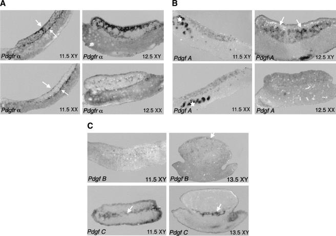
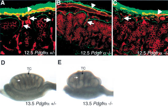
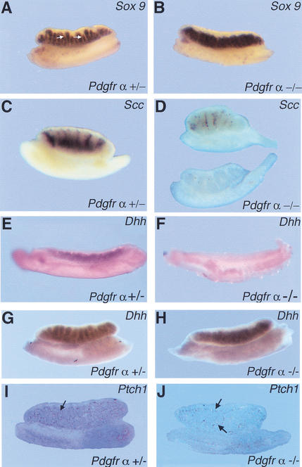
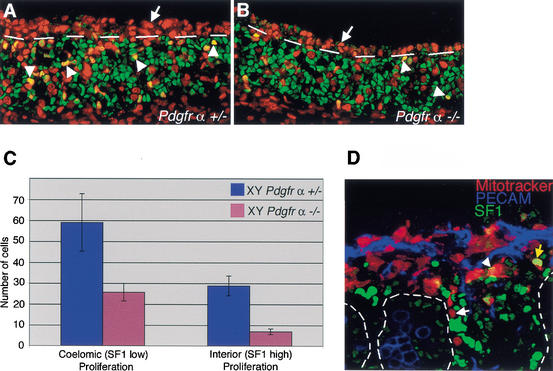
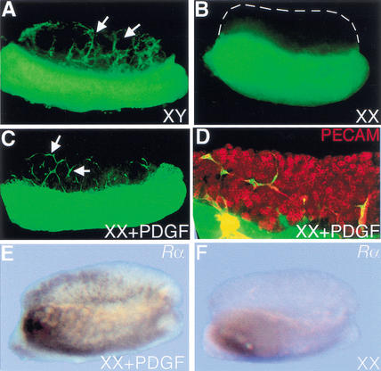
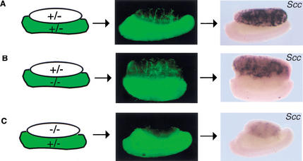
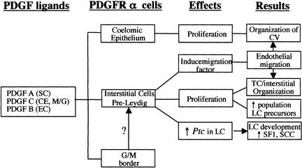
During testis development, the rapid morphological changes initiated by Sry require the coordinate integration of many signaling pathways. Based on the established role of the platelet-derived growth factor (PDGF) family of ligands and receptors in migration, proliferation, and differentiation of cells in various organ systems, we have investigated the role of PDGF in testis organogenesis. Analysis of expression patterns and characterization of the gonad phenotype in Pdgfr-alpha(-/-) embryos identified PDGFR-alpha as a critical mediator of signaling in the early testis at multiple steps of testis development. Pdgfr-alpha(-/-) XY gonads displayed disruptions in the organization of the vasculature and in the partitioning of interstitial and testis cord compartments. Closer examination revealed severe reductions in characteristic XY proliferation, mesonephric cell migration, and fetal Leydig cell differentiation. This work identifies PDGF signaling through the alpha receptor as an important event downstream of Sry in testis organogenesis and Leydig cell differentiation.


Figures

Figure 1
Figure 1
Expression of Pdgfr-α and Pdgf ligands at early stages of gonad development. In all figures anterior is to the left and the gonad is located on top of the mesonephros, positioned between the two white arrows as in A. (A) Pdgfr-α was expressed at the coelomic epithelium and mesonephros/gonad boundary (arrows) as well as on scattered cells in the interior of XY but not XX gonads. Expression was also detected at lower levels on mesenchymal cells of the mesonephros. Expression of the receptor was very strong in interstitial cells of the XY gonad at 12.5 dpc, but at much lower levels in the XX gonad. (B) Pdgf-A was expressed in both XY and XX gonads at 11.5 dpc as well as in the mesonephric tubules (*). High levels of Pdgf-A were detected inside the testis cords of XY gonads at 12.5 dpc (arrows), whereas expression was uniformly detected at low levels in 12.5-dpc XX gonads. (C) Pdgf-B is expressed in the gonad at low levels at 11.5 dpc. By 13.5 dpc, expression is exclusively endothelial, concentrated in the coelomic vessel of XY gonads (arrow). Pdgf-C expression is mainly concentrated at the mesonephros/gonad boundary of both XY gonads from 11.5 to 13.5 dpc (arrow).
Figure 2
Figure 2
Analysis of Pdgfr-α−/− testis structure at 12.5–13.5 dpc. (A) At 12.5 dpc, Pdgfr-α+/+ and Pdgfr-α+/− XY gonads have begun to form testis cords as outlined by laminin deposition (green). Staining with the endothelial and germ-cell marker PECAM (red) shows the formation of the coelomic vessel (arrowhead) and branches from the vessel that extend between testis cords (arrows). (B,C) In 12.5-dpc Pdgfr-α−/− XY samples, subdivision of testis cord and interstitial compartments had not occurred and the coelomic vessel (arrowhead) had formed few distinct branches between cords (arrows). (C) In a small proportion of samples, the coelomic vessel was not organized normally, and an increased number of endothelial cells was observed under the coelomic surface that were not organized as part of the coelomic vessel (red arrowhead). (D) By 13.5 dpc, testis cords (TC, black arrow) separated by regular interstitial spaces (white arrow) are well formed in Pdgfr-α+/− gonads. (E) At 13.5 dpc, Pdgfr-α−/− XY gonads had formed large, abnormally shaped testis cords (TC, black arrow) separated by irregular interstitial spaces (white arrow).
Figure 3
Figure 3
Expression of male-specific markers in Pdgfr-α−/− XY gonads. (A) Sox9 was expressed in the testis cords of Pdgfr-α+/− XY gonads at 12.5 dpc. Arrows point to interstitial space between testis cords in the Pdgfr-α+/− sample. (B) Sox9 was expressed in 12.5-dpc Pdgfr-α−/− XY gonads, but interstitial spaces between testis cords were absent. (C) Scc, a Leydig cell marker, is expressed extensively in the interstitium of Pdgfr-α+/− XY gonads at 13.5 dpc. (D) Scc was expressed at low levels or was absent in 13.5-dpc Pdgfr-α−/− XY gonads (two representative samples are shown). In 11.5-dpc Pdgfr-α−/− XY gonads, expression of Dhh is absent (F) compared with wild-type or heterozygous littermates (E). By 12.5 dpc, Dhh expression is expressed at normal levels in Pdgfr-α−/− XY gonads (H) compared with control XY gonads (G). (I) At 13.5 dpc, Ptch1 is normally expressed in interstitial cells (arrows). (J) In Pdgfr-α−/− XY gonads, Ptch1 expression is significantly reduced (arrows).
Figure 4
Figure 4
PDGFR-α is required for the proliferation of interstitial precursors at early stages of testis development. (A,B) At stages between 11.5 and 12.0 dpc, samples were stained with BrdU (red) to detect proliferating cells and SF1 (green) to stain Sertoli and Leydig cell precursors. (A) The coelomic epithelium of Pdgfr-α+/− XY gonads was highly proliferative in a population of cells that expressed low levels of SF1 (red cells above broken line, arrows). There were also many proliferating cells expressing SF1 at high levels within the gonad (arrowheads, yellow nuclei below broken line). (B) Pdgfr-α−/− XY gonads had 60% fewer proliferating cells at the coelomic epithelium (above broken line). SF1-positive proliferation within the gonad (arrowheads, below broken line) was also reduced by 70% compared with controls, as shown graphically in C. (C) Proliferation rates of SF1-low cells at the coelomic epithelium and SF1-high population in the gonad in Pdgfr-α+/− XY gonads (blue) and Pdgfr-α−/− XY gonads (pink). Error bars represent the standard deviation of the sample mean. (D) Mitotracker vital dye (red) was used to label cells at the coelomic epithelium at 22–23 ts in wild-type XY gonads. Samples were cultured for 40 h then stained with PECAM (blue) and SF1 (green). During culture, cells moved into the gonad, but only rarely expressed high levels of SF1, characteristic of Leydig cells (yellow arrow). Most cells derived from the coelomic epithelium were SF1 negative (white arrow) or SF1 low (arrowhead). Cord boundaries are outlined by dashed lines.
Figure 5
Figure 5
PDGFs induce endothelial cell migration into XX gonads. (A) XY gonads recruited cells (arrows) from GFP mesonephrons when recombinant cultures were assembled at 11.5 dpc and cultured for 48 h, whereas XX gonads did not (B, XX gonad outlined with broken lines). (C) When XX gonads were incubated in culture with 50 ng of PDGF-AA, PDGF-BB, or PDGF-AB, GFP-positive mesonephric cells were recruited into the gonad. (D) Following PDGF induction, samples were labeled with the endothelial and germ-cell marker, PECAM (red), which double-labeled all migrating cells and identified this population as endothelial. (E) Pdgfr-α was up-regulated in induced XX cultures compared with XX control cultures (F).
Figure 6
Figure 6
Analysis of migration and Leydig cell differentiation in Pdgfr-α−/− recombinant organ cultures. (A) At 12.5 dpc, Pdgfr-α+/+ or Pdgfr-α+/− XY gonads cultured with GFP-positive mesonephrons for 48 h recruited cells from the mesonephros (green, arrows). Following culture, Leydig cells differentiated as assayed by SCC expression. (B) When the gonadal portion of the culture was Pdgfr-α+/− and the mesonephros component was Pdgfr-α−/−, migration of mesonephric cells occurred normally (arrows), and Leydig cell development was normal compared with Pdgfr-α+/+ or Pdgfr-α+/− XY samples (A). (C) When the gonadal portion of the culture was Pdgfr-α−/−, cell migration (arrow) and Leydig cell development were both disrupted.
Figure 7
Figure 7
Effects of PDGFR-α signaling in the XY gonad. (A) Diagram of the expression of Pdgfr-α and its ligands and the cellular effects promoted by PDGF signaling in the XY gonad. CV, coelomic vessel; CE, coelomic epithelium; EC, endothelial cell; M/G, mesonephros/gonad border; SC, Sertoli cell; LC, Leydig cell.

References

    1. Ariyaratne S, Mendis-Handagama C, Hales B, Mason I. Studies on the onset of Leydig precursor cell differentiation in the prepubertal rat testis. Biol Reprod. 2000;63:165–171. - PubMed
    1. Betsholtz C, Karlsson L, Lindahlm P. Developmental roles of platelet-derived growth factors. Bioessays. 2001;23:494–507. - PubMed
    1. Bostrom H, Willetts K, Pekny M, Leveen P, Lindahl P, Hedstrand H, Pekna M, Hellstrom M, Gebre-Medhin S, Schalling M, et al. PDGF-A signaling is a critical event in lung alveolar myofibroblast development and alveogenesis. Cell. 1996;85:863–873. - PubMed
    1. Brennan J, Karl J, Capel B. Divergent vascular mechanisms downstream of Sry establish the arterial system in the XY gonad. Dev Biol. 2002;244:418–428. - PubMed
    1. Buehr M, Gu S, McLaren A. Mesonephric contribution to testis differentiation in the fetal mouse. Development. 1993;117:273–281. - PubMed

Publication types

MeSH terms

Substances