. 2003 Apr 15;548(Pt 2):373-82.
doi: 10.1113/jphysiol.2003.039131. Epub 2003 Mar 21.

GTP-induced tetrodotoxin-resistant Na+ current regulates excitability in mouse and rat small diameter sensory neurones

Affiliations

GTP-induced tetrodotoxin-resistant Na+ current regulates excitability in mouse and rat small diameter sensory neurones

Mark D Baker et al. J Physiol. .

Abstract

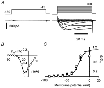
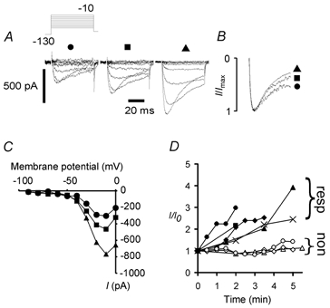
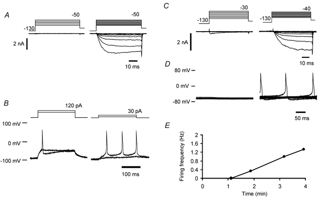
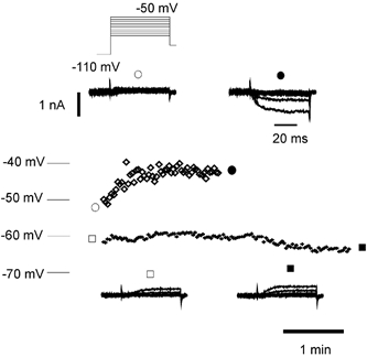
Peripheral pain thresholds are regulated by the actions of inflammatory mediators. Some act through G-protein-coupled receptors on voltage-gated sodium channels. We have found that a low-threshold, persistent tetrodotoxin-resistant Na+ current, attributed to NaV1.9, is upregulated by GTP and its non-hydrolysable analogue GTP-gamma-S, but not by GDP. Inclusion of GTP-gamma-S (500 microM) in the internal solution led to an increase in maximal current amplitude of > 300 % within 5 min. In current clamp, upregulation of persistent current was associated with a more negative threshold for action potential induction (by 15-16 mV) assessed from a holding potential of -90 mV. This was not seen in neurones without the low-threshold current or with internal GDP (P < 0.001). In addition, persistent current upregulation depolarized neurones. At -60 mV, internal GTP-gamma-S led to the generation of spontaneous activity in initially silent neurones only when persistent current was upregulated. These findings suggest that regulation of the persistent current has important consequences for nociceptor excitability.


Figures

Figure 3
Figure 3. Upregulation of persistent current in a NaV1.8 null mutant neurone following exposure to 500 μm GTP-γ-S
A: left, maximal inward current recorded at −15 mV as soon as possible following membrane rupture. Right, persistent current family evoked by incrementing voltage-clamp steps 12 min later. B, peak current (I) versus membrane potential (Vm) relation for the GTP-γ-S-upregulated current in A. Current activated at potentials more negative than −50 mV. C, normalized peak conductance versus membrane potential plot for the same neurone as in A and B (○). Also shown are normalized peak conductance versus membrane potential plots for the neurone in Fig. 1A (symbols for 0, 2 and 7 min: •, ▪, and ▴, respectively). The data are normalized to conductance values at −10 mV because the increasing rate of inactivation and residual K+ currents at positive potentials begin to limit inward current amplitudes at early times. The smooth curve fitted to data at 7 min (▴) is a Boltzmann function drawn with best-fit parameters (V1/2 = −23.8 mV, ag = 6.85 mV). All conductances were calculated assuming ENa = +45 mV.
Figure 1
Figure 1. GTP increases the amplitude of persistent Na+ current in NaV1.8 null mutant neurones
A, with 500 μm GTP in the internal solution the amplitude of low-threshold current increased dramatically over 7 min by more than 100 % (from left: 0, 2, 7 min). The voltage-clamp protocol is shown in the inset (mV). B, normalized peak currents at 0, 2 and 7 min (•, ▪, ▴, respectively) indicating no major changes in current activation kinetics and a slight increase in the amount of inactivation with time. C, the voltage dependence of current activation was essentially unchanged despite the large increase in current amplitude from 0 to 7 min. D, 5 of 8 neurones exhibited substantial upregulation over the first 5 min of voltage clamp. The 5 neurones responding (resp) are represented by •, ▪, ♦, ▴ and ×; the 3 non-responding (non) neurones are represented by open symbols.
Figure 4
Figure 4. Measurement of voltage threshold and GTP-γ-S-related fall in voltage threshold for action potential induction
A, depolarizing currents of 200 and 300 pA applied to an example wild-type neurone gave rise to subthreshold electrotonic responses. Thick curves are exponentials drawn according to best-fit parameters derived from the initial portion of the response. An action potential was elicited by 400 pA. B, internal GTP-γ-S in a NaV1.8 null mutant neurone gave rise to a reduction in voltage threshold (tested from a holding potential of −90 mV), and prolonged applied depolarizations. The reduction in threshold took place over 13 min with intracellular GTP-γ-S. Enhanced low threshold current gave rise to profound prolongation of previously subthreshold depolarizations (arrow), consistent with a substantial increase in low-threshold current amplitude and slow current kinetics (before and after threshold reduction, left and right panels, respectively). C, both NaV1.8 null mutant and wild-type (WT) dorsal root ganglion neurones exhibited falls in threshold when persistent inward current was upregulated by internal GTP-γ-S (□, n = 3 for both NaV1.8 null and wild-type; means ±s.e.m.), but where no persistent current could be recorded, the threshold increased slightly (▪, n = 16 and 11 for Nav1.8 null and wild-type, respectively; means ±s.e.m., P < 0.0001, Student's two-tailed unpaired t test, pooled data), strongly implying that the presence of the current gave rise to the threshold fall. No such fall in voltage threshold was found in 15 neurones with internal GDP.
Figure 5
Figure 5. Persistent current upregulation alters the voltage threshold and leads to accommodation breakdown
A, voltage-clamp recordings using quasi-physiological solutions from a wild-type neurone before and after persistent current upregulation (left and right panels, respectively). Persistent current began to activate over a potential range that was more negative than the threshold for K+ current recruitment. B, responses to 200 ms duration ‘just‘ subthreshold and supra-threshold depolarizations from a holding potential of −90 mV, before (left) and after (right) current upregulation. Upregulation of persistent current lowered voltage and current threshold and gave rise to repetitive firing (same neurone as in A). Repetitive firing occurred in this neurone at a frequency of 13 Hz, where the most negative potential during the interspike interval was −77 mV. C, voltage-clamp recordings using quasi-physiological solutions in an example wild-type neurone. At the start of recording a transient Na+ current began to activate at −30 mV (left). After upregulation of NaV1.9, a persistent current appeared (right). D, current-clamp recordings showing 10 sequentially recorded traces at 1 min (left) and 3 min (right) after the whole-cell configuration had been achieved (same neurone as in C). The holding current was not greater than −20 pA, and was adjusted to maintain membrane potential at close to −60 mV. E, example of the time dependence of discharge frequency. Same neurone as in C and D.
Figure 2
Figure 2. Internal GTP-γ-S but not GDP leads to substantial persistent current upregulation in Nav1.8 null mutant neurones
A, internal GTP-γ-S (500 μm) consistently increased maximal current amplitude on average by > 300 % over 5 min (•, n = 7, means ±s.e.m.). In neurones from NaV1.8 null mutants, current amplitude did not increase with intracellular GDP (500 μm) for recordings over 2–40 min (found in a total of 10 neurones); data for 4 neurones are shown (○, means ±s.e.m.). B, with H7 (100 μm), ATP (3 mm) and GTP-γ-S in the internal solution, the mean current increased in amplitude (♦, n = 7, means +s.e.m.), but with H7 (200 μm) and GTP-γ-S only in the pipette solution the upregulation was significantly reduced (⋄, n = 5, means - s.e.m.; ANOVA, P < 0.001). Some s.e.m. values were smaller than the symbol size. C, internal solution containing 2.5 mm Li+ did not cause upregulation of the persistent current in 4 neurones (•, ▪, ♦, ▴; mean data plotted as ○). D, Li+ (2.5 mm) with GDP (500 μm) also did not induce upregulation of the current (n = 3; ▪, ♦, individual neurones; mean data ±s.e.m. plotted as ○).
Figure 6
Figure 6. Upregulation of persistent current by GTP-γ-S affects membrane potential
Top, upregulation of persistent current by GTP-γ-S depolarized a wild-type neurone. Membrane potential recorded in current clamp plotted over 2 min is shown (⋄), with voltage-clamp recordings (inset) from the same neurone before (○) and after (•) current upregulation. Input conductance fell from 6.96 to 5.93 nS over the same period. Bottom, in another wild-type neurone (+), there was no current upregulation over 4 min and no depolarization. Inset, voltage-clamp records from the same neurone plotted before (□) and after (▪) membrane potential recording. The calibration bars in the top inset also apply to the bottom inset.

References

    1. Akopian AN, Okuse K, Souslova V, England S, Ogata N, Wood JN. Trans-splicing of a voltage-gated sodium channel is regulated by nerve growth factor. FEBS Lett. 1999a;445:177–182. - PubMed
    1. Akopian AN, Souslova V, England S, Okuse K, Ogata N, Ure J, Smith A, Kerr BJ, McMahon SB, Boyce S, Hill R, Stanfa LC, Dickenson AH, Wood JN. The tetrodotoxin-resistant sodium channel SNS has a specialized function in pain pathways. Nat Neurosci. 1999b;2:541–548. - PubMed
    1. Alroy G, Su H, Yaari Y. Protein kinase C mediates muscarinic block of intrinsic bursting in rat hippocampal neurons. J Physiol. 1999;518:71–79. - PMC - PubMed
    1. Amaya F, Decosterd I, Samad TA, Plumpton C, Tate S, Mannion RJ, Costigan M, Woolf CJ. Diversity of expression of the sensory neuron-specific TTX-resistant voltage-gated sodium ion channels SNS and SNS2. Mol Cell Neurosci. 2000;15:331–342. - PubMed
    1. Baker MD, Bostock H. Low-threshold, persistent sodium current in rat large dorsal root ganglion neurons in culture. J Neurophys. 1997;77:1503–1513. - PubMed

Publication types

MeSH terms

LinkOut - more resources