. 2003 May 15;549(Pt 1):3-19.
doi: 10.1113/jphysiol.2003.039818. Epub 2003 Mar 21.

Acute regulation of the SLC26A3 congenital chloride diarrhoea anion exchanger (DRA) expressed in Xenopus oocytes

Affiliations

Acute regulation of the SLC26A3 congenital chloride diarrhoea anion exchanger (DRA) expressed in Xenopus oocytes

Marina N Chernova et al. J Physiol. .

Abstract

Mutations in the human SLC26A3 gene, also known as down-regulated in adenoma (hDRA), cause autosomal recessive congenital chloride-losing diarrhoea (CLD). hDRA expressed in Xenopus oocytes mediated bidirectional Cl--Cl- and Cl--HCO3- exchange. In contrast, transport of oxalate was low, and transport of sulfate and of butyrate was undetectable. Two CLD missense disease mutants of hDRA were nonfunctional in oocytes. Truncation of up to 44 C-terminal amino acids from the putatively cytoplasmic C-terminal hydrophilic domain left transport function unimpaired, but deletion of the adjacent STAS (sulfate transporter anti-sigma factor antagonist) domain abolished function. hDRA-mediated Cl- transport was insensitive to changing extracellular pH, but was inhibited by intracellular acidification and activated by NH4+ at acidifying concentrations. These regulatory responses did not require the presence of either hDRA's N-terminal cytoplasmic tail or its 44 C-terminal amino acids, but they did require more proximate residues of the C-terminal cytoplasmic domain. Although only weakly sensitive to inhibition by stilbenes, hDRA was inhibited with two orders of magnitude greater potency by the anti-inflammatory drugs niflumate and tenidap. cAMP-insensitive Cl--HCO3- exchange mediated by hDRA gained modest cAMP sensitivity when co-expressed with cystic fibrosis transmembrane conductance regulator (CFTR). Despite the absence of hDRA transcripts in human cell lines derived from CFTR patients, DRA mRNA was present at wild-type levels in proximal colon and nearly so in the distal ileum of CFTR(-/-) mice. Thus, pharmacological modulation of DRA might be a useful adjunct treatment of cystic fibrosis.


Figures

Figure 1
Figure 1. Anion selectivity of human down-regulated in adenoma (hDRA)-mediated isotopic fluxes
A, hDRA-mediated 36Cl influx; representative of 6 similar experiments. B, [14C]butyrate influx into oocytes pre-injected with water or with hDRA cRNA; one of two similar experiments. C, hDRA-mediated [14C]oxalate influx; representative of three similar experiments (*P < 0.05). D, [35S]sulfate influx into oocytes preinjected with water or with hDRA cRNA; one of two similar experiments (see opposite). The number of oocytes tested is given in parentheses. E, [35S]sulfate efflux from DRA-expressing oocytes is not activated by acid bath pH. Efflux was measured from 5 individual oocytes each transferred from Cl-free bath into pH 5 ND-96 and then into pH 7.4 ND-96. Inset: [35S]sulfate efflux rate constants in each condition. F, 36Cl efflux from two water-injected oocytes (upper traces) and from 5 DRA-expressing oocytes (lower traces) first in the absence of Cl, then transferredinto 64 mm sulfate, then in ND-96 and finally returned to the Cl-free bath. Inset: 36Cl efflux rate constants for DRA-expressing oocytes.
Figure 3
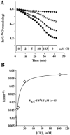
Figure 3. Extracellular Cl dependence of hDRA-mediated 36Cl efflux
A, 36Cl efflux into bath solutions containing increasing Cl concentrations. Uppermost trace represents a water-injected oocyte. Lower traces represent three hDRA-expressing oocytes. B, [Cl] dependence of hDRA-mediated 36Cl efflux from a single representative oocyte. The curve is the best Michaelis-Menten fit to the data. Mean K1/2(Clo) was 5.8 ± 1.5 mm for 13 hDRA-expressing oocytes studied with the same protocol.
Figure 4
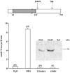
Figure 4. CLD mutations in STAS domain exhibit loss of function
Top: schematic diagram showing location of the chloride-losing diarrhoea (CLD) missense mutations I544N, and 2116delA which leads to termination at codon 711. Open bars indicate hydrophilic, putative cytoplasmic domains. The grey bar indicates the hydrophobic polytopic transmembrane domain. Hatch marks indicate that grey bar is shorter than if it was drawn to scale. Amino acid residue numbers under the bar mark approximate domain boundaries. Bottom: 36Cl influx assay shows that both missense mutants exhibit loss of function; numbers of oocytes tested given in parentheses. Injected cRNA was 1 ng for wild-type (WT) hDRA and 10 ng for each mutant. Increasing cRNA to 50 ng did not lead to 36Cl uptake by the mutants. Inset: hDRA immunoblot of whole oocyte lysates comparing WT hDRA with hDRA I544N polypeptide (10 ng injected cRNA for each). hDRA 2116delA, lacking the C-terminus, is undetectable by the anti-hDRA C-terminal peptide antibody.
Figure 5
Figure 5. Effects of cytoplasmic domain truncations on hDRA basal activity
A, left, schematic diagram showing C-terminal region of hDRA across which C-terminal deletions were generated. Italics indicate consensus binding site for PDZ domain. Asterisks indicate potential phosphoryation sites. A, right, 36Cl efflux rate constants exhibited by WT hDRA and by the indicated hDRA mutants. One nanogram of cRNA was injected for all constructs. * Y756F differs significantly from WT DRA and all other groups by ANOVA. B, left, schematic diagram depicting the more extensive deletions generated. B, right, 36Cl efflux rate constants exhibited by WT hDRA and by the indicated hDRA mutants. One nanogram of cRNA was injected for WT hDRA and 720X; 10 ng cRNA injected for the mutants (-STAS), 524X and ΔN52. Number of oocytes tested given in parentheses. * Significant difference from WT DRA by ANOVA. Inset, hDRA STAS domain deletion polypeptide accumulates in oocytes to higher-than-WT levels. hDRA 524X cannot be detected by the anti-hDRA C-terminal peptide antibody.
Figure 2
Figure 2. hDRA mediates Cl-Cl and Cl-HCO3 exchange
A, 36Cl efflux traces from water-injected oocytes (upper three traces) and from hDRA-expressing oocytes (lower four traces) during sequential incubations in baths without Cl, baths containing 24 mm HCO3 and baths containing 96 mm Cl (ND-96). B, relative increase in stimulation of 36Cl efflux upon changing from Cl-free baths to baths containing the indicated anion (n = 4). C, pHi of representative oocytes previously injected with water (□) or with hDRA cRNA (♦) during bath Cl removal and restoration. pHi was measured by 2′,7′-bis(carboxyethyl)-5-(and −6)-carboxyfluorescein (BCECF) fluorescence ratio imaging. D, upper panel: pHi of representative oocytes previously injected with water (lower trace) or with hDRA cRNA (upper trace) during bath Cl removal and restoration. pHi was measured by pH-sensitive microelectrode. D, lower panel: membrane potential (Vm) traces recorded from the same oocytes previously injected with water (upper trace) or with hDRA cRNA (lower trace).
Figure 6
Figure 6. hDRA is inhibited by intracellular, but not by extracellular acidification
A, 36Cl efflux from a water-injected oocyte (uppermost trace) and several hDRA-expressing oocytes (lower traces) during transition from bath pH 5.0 to 7.4, followed by exposure to 2 mm DIDS at pH 7.4. B, 36Cl efflux from a water-injected oocyte (uppermost trace) and several oocytes expressing either hDRA 524X (middle traces) or WT hDRA (lower traces) during exposure and removal of 40 mm butyrate, followed by switch to Cl-free bath. C, summary of effect of pHo on WT hDRA-mediated 36Cl efflux. D, summary of relative increase in activation by butyrate removal of 36Cl efflux from oocytes expressing WT hDRA or the indicated mutants. Number of oocytes tested given in parentheses.
Figure 7
Figure 7. hDRA is stimulated by NH4+
A, 36Cl efflux from a water-injected oocyte (uppermost trace) and several oocytes expressing either hDRA 524X (middle traces) or WT hDRA (lower traces) during exposure to NH4+, followed by transition to Cl-free bath. B, relative increase in stimulation by NH4+ of hDRA-mediated 36Cl efflux from oocytes expressing either WT hDRA or the indicated mutant polypeptides; *P < 0.05 by ANOVA. Number of oocytes tested given in parentheses.
Figure 8
Figure 8. hDRA is inhibited by tenidap much more potently than by stilbenes
A, 36Cl efflux into bath solutions containing increasing tenidap concentrations. Uppermost trace represents a water-injected oocyte. Lower traces represent three hDRA-expressing oocytes. B, tenidap concentration dependence of hDRA-mediated 36Cl efflux from a single oocyte. Curve is the best fit to this data of the Michaelis-Menten equation. Mean ID50 for tenidap for 6 similar hDRA-expressing oocytes was 9.6 ± 1.9 μm.
Figure 9
Figure 9. CFTR co-expression confers modest cAMP-sensitivity upon hDRA-mediated Cl-HCO3 exchange
A, hDRA-mediated Cl-HCO3 exchange activity measured by BCECF fluorescence ratio imaging in representative oocytes subjected to sequential Cl removal and restoration in the absence (○) or presence (•) of 2 mm cAMP plus 1 mm IBMX. B, Cl-HCO3 exchange activity in representative oocytes expressing CFTR and subjected to sequential Cl removal and restoration in the absence (▵) or presence (▴) of 2 mm cAMP plus 1 mm IBMX. C, Cl-HCO3 exchange activity in representative oocytes co-expressing hDRA and CFTR, subjected to sequential Cl removal and restoration in the absence (▵) or presence (▴) of 2 mm cAMP plus 1 mm IBMX. D, summarized results of Cl-HCO3 exchange activity for the number of oocytes given in parentheses. Resting pHi was 7.03 ± 0.02 for ‘H2O’ and ‘DRA’ (minus cAMP) groups, and 7.12 ± 0.02 for all other groups. Note that stimulation by cAMP and IBMX is observed only in oocytes co-expressing CFTR and hDRA3. *P < 0.001 compared to water-injected oocytes; **P < 0.001 compared to absence of cAMP/IBMX (adjacent bar).
Figure 10
Figure 10. DRA mRNA levels in CFTR(−/−) and (+/+) mice
RNAse protection assays of mouse DRA mRNA in whole proximal colon (caecum, A and C) and in distal ileum (B and D) from individual CFTR(+/+), (−/−) and (−/+) mice. Replica digestions of RNA from colon and ileum were tested for each numbered individual. Experiments 1 (A and B) and 2 (C and D) represent separate groups of age-matched individuals. Experiment 2 is representative of three independent digestions with the same RNA samples. The 18S and 28S RNAs were intact in all samples (not shown). 32P-end-labelled size standards mark 300 and 400 nt. S, probe protected by hybridization with unlabelled sense DRA cRNA; A, probe unprotected by hybridization with unlabelled antisense DRA cRNA.

References

    1. Aichbichler BW, Zerr CH, Santa Ana CA, Porter JL, Fordtran JS. Proton-pump inhibition of gastric chloride secretion in congenital chloridorrhea. N Engl J Med. 1997;336:106–109. - PubMed
    1. Alper SL. Genetic diseases of acid-base transport. Annu Rev Physiol. 2002;64:899–923. - PubMed
    1. Antalis TM, Reeder JA, Gotley DC, Byeon MK, Walsh MD, Henderson KW, Papas TS, Schweinfest CA. Down-regulation of the Down-Regulated in Adenoma (DRA) gene correlates with colon tumor progression. Clin Cancer Res. 1998;4:1857–1863. - PubMed
    1. Aravind L, Koonin EV. The STAS domain-a link between anion transporters and antisigma-factor antagonists. Curr Biol. 2000;10:R53, R55. - PubMed
    1. Bennekou P, Pedersen O, Moller A, Christophersen P. Volume control in sickle cells is facilitated by the novel anion conductance inhibitor NS1652. Blood. 2000;5:1842–1848. - PubMed

Publication types

MeSH terms

LinkOut - more resources