. 2003 Apr 1;100(7):4102-7.
doi: 10.1073/pnas.0730835100. Epub 2003 Mar 21.

Activation-induced cytidine deaminase deaminates deoxycytidine on single-stranded DNA but requires the action of RNase

Affiliations

Activation-induced cytidine deaminase deaminates deoxycytidine on single-stranded DNA but requires the action of RNase

Ronda Bransteitter et al. Proc Natl Acad Sci U S A. .

Abstract

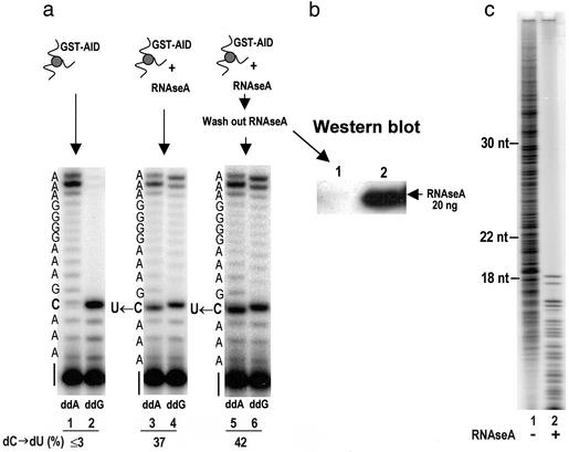
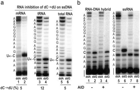
The expression of activation-induced cytidine deaminase (AID) is prerequisite to a "trifecta" of key molecular events in B cells: class-switch recombination and somatic hypermutation in humans and mice and gene conversion in chickens. Although this critically important enzyme shares common sequence motifs with apolipoprotein B mRNA-editing enzyme, and exhibits deaminase activity on free deoxycytidine in solution, it has not been shown to act on either RNA or DNA. Recent mutagenesis data in Escherichia coli suggest that AID may deaminate dC on DNA, but its putative biochemical activities on either DNA or RNA remained a mystery. Here, we show that AID catalyzes deamination of dC residues on single-stranded DNA in vitro but not on double-stranded DNA, RNA-DNA hybrids, or RNA. Remarkably, it has no measurable deaminase activity on single-stranded DNA unless pretreated with RNase to remove inhibitory RNA bound to AID. AID catalyzes dC --> dU deamination activity most avidly on double-stranded DNA substrates containing a small "transcription-like" single-stranded DNA bubble, suggesting a targeting mechanism for this enigmatic enzyme during somatic hypermutation.


Figures

Figure 1
Figure 1
AID-catalyzed dC → dU conversion on ssDNA potentiated by RNase. (a) Assay 1 detects dC deamination by using UDG and APE. An AID-catalyzed 14-nt deamination product occurs with ssDNA but only in the presence of RNaseA (lane 1). (b) dC → dU conversion in SHM hot-spot and non-hot-spot sequence contexts using assay 1. The arrows indicate positions of deamination products after UDG and APE treatment. (c) Assay 2 detects dC deamination by using primer elongation–dideoxynucleotide termination. Deamination of dC is detected by the presence of an intense termination band (in lane 1, ddA) located opposite the original template C site (U ← C, see sequence to the left of gel) and a reduction in the intensity of the termination band (in lane 2, ddG) located opposite the original template C site. Concomitant with the conversion of dC → dU is the presence of bands migrating past the U ← C template site (lane 2), denoted by the bracket to the right of the gel.
Figure 2
Figure 2
RNase activates AID by digesting AID-associated inhibitor RNA. (a) RNase pretreatment of AID is sufficient to observe AID-catalyzed dC → dU conversion on ssDNA as detected by primer elongation–dideoxynucleotide termination (assay 2). GST-AID bound to glutathione-Sepharose beads was preincubated with RNaseA for 5 min at 37°C and washed extensively to remove the RNaseA. AID-catalyzed dC → dU conversion after RNaseA removal can be observed in lanes 5 and 6 (U ← C template site, indicated at the left of the gel). The fraction of dC → dU conversion is indicated at the bottom of the gel as dC → dU (%). (b) Western blot showing the efficacy of RNaseA removal by washing the GST-AID-bound beads indicated by the absence of a crossreacting band with RNaseA antibody (lane 1). (c) Detection of AID-associated inhibitor RNA. After incubation of AID with proteinase K, a phenol/chloroform/isoamyl extraction was carried out followed by 5′-32P labeling of putative nucleic acids by using T4 polynucleotide kinase and resolution of labeled products by 20% denaturing PAGE. The absence of bands >18 nt in the RNase-treated sample (lane 2) demonstrates the existence of AID-associated RNA. The appearance of bands <18 nt in the RNase-treated sample (lane 2) represents RNase-digested products; RNaseA hydrolyzes ssRNA at C and U sites. Size markers at the left indicate the position to which DNA oligonucleotides have migrated on the gel.
Figure 3
Figure 3
Activity of RNase-activated AID attached to GST-Sepharose beads. (a) Inhibition of AID-catalyzed dC → dU deamination on ssDNA substrate by exogenous RNA. AID attached to GST resin was activated by RNaseA as described for Fig. 2. Activated AID beads (25 μl) were preincubated with 1.25 μg of mRNA (from Ramos cells), tRNA (E. coli), or total RNA (heart cells) for 3 min at 37°C and used in the deamination reaction with ssDNA substrate. The fraction of dC → dU conversion is indicated at the bottom of the gel as dC → dU (%). (b) AID does not deaminate dC in RNA–DNA hybrid or C in ssRNA. RNA–DNA hybrid or ssRNA was incubated in the reaction buffer at 37°C for 5 min in the presence or absence of activated AID-GST beads. The deamination product for ssRNA was analyzed by assay 2, in which avian myeloblastosis virus reverse transcriptase was used instead of T7 sequenase.
Figure 4
Figure 4
AID-deamination activities. (a) Time course for AID (200 nM)-catalyzed dC → dU conversion on ssDNA (100 nM) in the presence of 1 μg of RNaseA (filled circles) or 0.1 μg of RNaseA, for which either RNase inhibitor was absent (open circles) or absent at t = 0 and added to the reaction at t = 5 min (see arrow) (filled triangles). (b) AID-catalyzed deamination rates as a function of AID concentration for various cytosine-containing substrates. Filled circles, dC → dU on ssDNA; filled triangles, 5-MeC → T on ssDNA (in 5′-CpG sequence context); open circles, 5-MeC → T on ssDNA (in non-5′-CpG sequence context). (Inset) AID-catalyzed deamination rates as a function of AID concentration for the free deoxynucleoside, CdR (open circles), and free nucleoside, CR (filled circles), in the absence of RNase.

References

    1. Muramatsu M, Kinoshita K, Fagarasan S, Yamada S, Shinkai Y, Honjo T. Cell. 2000;102:553–563. - PubMed
    1. Revy P, Muto T, Levy Y, Geissmann F, Plebani A, Sanal O, Catalan N, Forveille M, Dufourcq-Labelouse R, Gennery A, et al. Cell. 2000;102:565–575. - PubMed
    1. Martin A, Bardwell P D, Woo C J, Fan M, Shulman M J, Scharff M D. Nature. 2002;415:802–806. - PubMed
    1. Martin A, Scharff M D. Proc Natl Acad Sci USA. 2002;99:12304–12308. - PMC - PubMed
    1. Yoshikawa K, Okazaki I M, Eto T, Kinoshita K, Muramatsu M, Nagaoka H, Honjo T. Science. 2002;296:2033–2036. - PubMed

Publication types