. 2003 Apr 1;100(7):3901-6.
doi: 10.1073/pnas.0630165100. Epub 2003 Mar 21.

Genetic characterization of glucose transporter function in Leishmania mexicana

Affiliations

Genetic characterization of glucose transporter function in Leishmania mexicana

Richard J S Burchmore et al. Proc Natl Acad Sci U S A. .

Erratum in

  • Proc Natl Acad Sci U S A. 2003 May 13;100(10):6287

Abstract

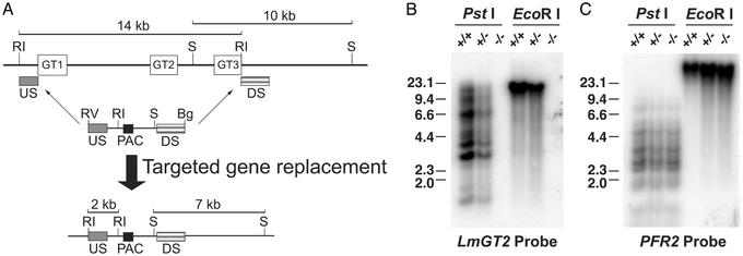
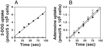
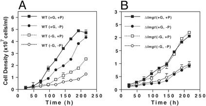
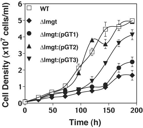
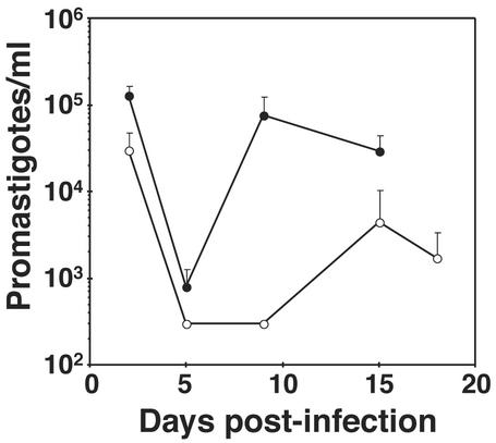
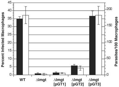
Both insect and mammalian life cycle stages of Leishmania mexicana take up glucose and express all three isoforms encoded by the LmGT glucose transporter gene family. To evaluate glucose transporter function in intact parasites, a null mutant line has been created by targeted disruption of the LmGT locus that encompasses the LmGT1, LmGT2, and LmGT3 genes. This deltalmgt null mutant exhibited no detectable glucose transport activity. The growth rate of the deltalmgt knockout in the promastigote stage was reduced to a rate comparable with that of WT cells grown in the absence of glucose. deltalmgt cells also exhibited dramatically reduced infectivity to macrophages, demonstrating that expression of LmGT isoforms is essential for viability of amastigotes. Furthermore, WT L. mexicana were not able to grow as axenic culture form amastigotes if glucose was withdrawn from the medium, implying that glucose is an essential nutrient in this life cycle stage. Expression of either LmGT2 or LmGT3, but not of LmGT1, in deltalmgt null mutants significantly restored growth as promastigotes, but only LmGT3 expression substantially rescued amastigote growth in macrophages. Subcellular localization of the three isoforms was investigated in deltalmgt cells expressing individual LmGT isoforms. Using anti-LmGT antiserum and GFP-tagged LmGT fusion proteins, LmGT2 and LmGT3 were localized to the cell body, whereas LmGT1 was localized specifically to the flagellum. These results establish that each glucose transporter isoform has distinct biological functions in the parasite.


Figures

Figure 1
Figure 1
Construction of the glucose transporter null mutant, Δlmgt, by targeted gene replacement. (A) Strategy for targeted gene replacement. (Upper) The LmGT gene locus including the three ORFs (open rectangles marked GT1, GT2, and GT3), the 10-kb SmaI and 14-kb EcoRI restriction fragments, and the location of the upstream (US) and downstream (DS) segments used to target homologous recombination of the gene disruption constructs. One of these disruption constructs is shown immediately below the LmGT locus and includes the US and DS segments, the ORF for the PAC selectable marker, the EcoRV and BglII terminal polylinker restriction sites, and the internal EcoRI and SmaI restriction sites. The thin arrows indicate the sites of homologous integration. (Lower) The targeted gene replacement event is indicated below the thick arrow, showing the structure of the resulting chromosomal locus and the predicted 2-kb EcoRI and 7-kb SmaI restriction fragments that are diagnostic of the correct homologous integration event. Symbols indicating restriction fragments are: RI, EcoRI; S, SmaI; RV, EcoRV; and Bg, BglII. Generation of the null mutant required a second targeted gene replacement using a similar gene disruption cassette containing a SAT marker. (B) Southern blot containing 10 μg of genomic DNA from WT parasites (+/+), heterozygous knockout line after integration of the PAC gene disruption construct (+/−), or Δlmgt null mutant (−/−) digested with PstI or with EcoRI and hybridized with a radiolabeled probe representing the LmGT2 ORF. The numbers indicate the positions and sizes (kilobase pairs) of DNA molecular weight markers. (C) The same blot shown in B after elution of the LmGT2 probe and rehybridization to the PFR2 probe that encodes an unrelated paraflagellar rod protein.
Figure 2
Figure 2
Uptake of [3H]2-deoxy-d-glucose (2-DOG, A) and [3H]adenosine (B) by WT (filled circles) and Δlmgt null mutant (open circles) parasites. The error bars in this and other figures indicate the SD.
Figure 3
Figure 3
Growth of WT (A) and Δlmgt (B) promastigotes in DME-L medium containing (+) or lacking (−) 25 mM glucose (G) or 5 mM proline (P). Parasites were inoculated at an initial density of 1 × 10⁶ cells per ml, and aliquots of the cultures were counted (n = 3, average ± SD) on a hemacytometer at various times thereafter.
Figure 4
Figure 4
Growth in RPMI medium of promastigotes of WT, Δlmgt null mutant, and Δlmgt null mutants complemented with each of the LmGT genes (Δlmgt[pGT1], Δlmgt[pGT2], Δlmgt[pGT3]).
Figure 5
Figure 5
Infection of L. longipalpis sandflies by WT (filled circles) and Δlmgt (open circles) promastigotes. At each time point after infection, midguts were dissected from 10–12 sandflies, and parasites were quantitated. Similar results were obtained from four independent experiments.
Figure 6
Figure 6
Growth of WT, Δlmgt, Δlmgt[pGT1], Δlmgt[pGT2], and Δlmgt[pGT3] lines in murine peritoneal macrophages. Filled bars represent percent of infected macrophages, and open bars represent parasites per 100 macrophages for the same fields. Primary peritoneal macrophages were infected with stationary phase promastigotes, and the number of intracellular amastigotes (n = 3, average ± SD) was quantitated 6 days after infection.
Figure 7
Figure 7
Growth of WT L. mexicana and Δlmgt null mutants as axenic CF amastigotes. (A) WT (circles) and Δlmgt (triangles) parasites were inoculated into Schneider's medium supplemented with 20% iFCS and adjusted to pH 5.5 followed by incubation at 32.5°C, and aliquots were withdrawn and counted (n = 3, average ± SD) at various times. (B) CF amastigotes of WT L. mexicana, growing in DME-L medium containing 30 mM Mes buffer, pH 5.5, and supplemented with 20% iFCS (CF-DME-L), were pelleted, washed, and inoculated at a density of 1 × 10⁶ cells per ml into fresh CF-DME-L constituted with 11 mM glucose and dialyzed iFCS (diamonds), or CF-DME-L deficient in glucose and constituted with dialyzed iFCS (squares).

References

    1. Anez N, Luga A, Loaiza A, Nieves D, Orozco J. Med Vet Entomol. 1994;8:38–42. - PubMed
    1. Sacks D L, Perkins P V. Science. 1984;223:1417–1419. - PubMed
    1. Schlein Y. Parasitol Today. 1986;2:175–177. - PubMed
    1. Tang Y, Ward R D. Med Vet Entomol. 1988;12:13–19. - PubMed
    1. Richards A G. Acta Trop. 1975;32:83–95. - PubMed

Publication types

Substances