

Activity-dependent regulation of the subcellular localization of neuronal calcium sensor-1 in the avian cochlear nucleus
- PMID: 12654347
- PMCID: PMC1847351
- DOI: 10.1016/s0306-4522(02)00928-4
Activity-dependent regulation of the subcellular localization of neuronal calcium sensor-1 in the avian cochlear nucleus
Abstract
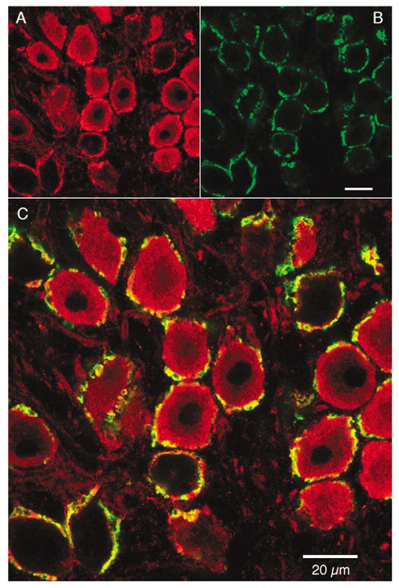
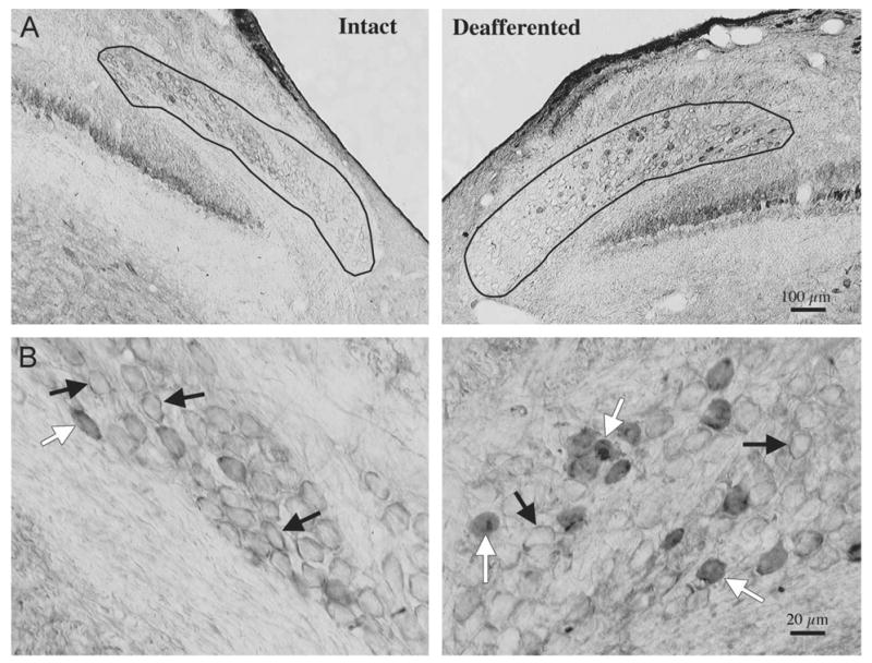
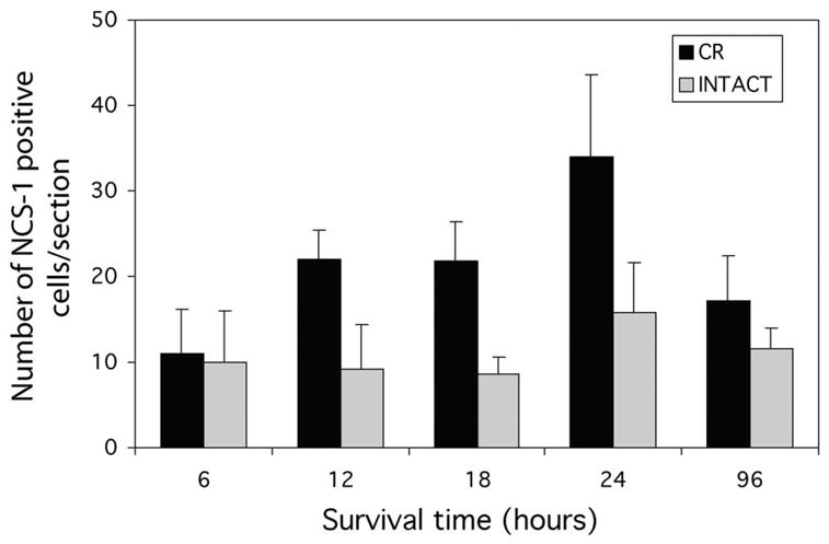
Neurons in the avian cochlear nucleus, nucleus magnocellularis (NM), are highly sensitive to manipulations of afferent input, and removal of afferent activity through cochlear ablation results in the death of approximately 20-40% of ipsilateral NM neurons. The intracellular cascades that determine whether an individual NM neuron will die or survive are not fully understood. One early event observed in NM following deafferentation is a rapid rise in intracellular calcium concentration. In most cellular systems, the activity of calcium-binding proteins is believed to accommodate calcium influx. The calcium-binding protein, neuronal calcium sensor-1 (NCS-1), is an intracellular neuronal calcium sensor belonging to the EF-hand superfamily. NCS-1 has been implicated in calcium-dependent regulation of signaling cascades. To evaluate NCS-1 action in NM neurons, the localization of NCS-1 protein was examined. Double-label immunofluorescence experiments revealed that NCS-1 expression is evident in both the presynaptic nerve terminal and postsynaptic NM neuron. The postsynaptic expression of NCS-1 typically appears to be closely associated with the cell membrane. This close proximity of NCS-1 to the postsynaptic membrane could allow NCS-1 to function as a modulator of postsynaptic signaling events. Following deafferentation, NM neurons were more likely to show diffuse cytoplasmic NCS-1 labeling. This increase in the number of cells showing diffuse cytoplasmic labeling was observed 12 and 24 h following cochlea ablation, but was not observed 4 days following surgery. This activity-dependent regulation of NCS-1 subcellular localization suggests it may be associated with, or influenced by, processes important for the survival of NM neurons.
Figures
References
-
- Baimbridge KG, Celio MR, Rogers JH. Calcium-binding proteins in the nervous system. Trends Neurosci. 1992;15:303–308. - PubMed
-
- Bartlett SE, Reynolds AJ, Weible M, Jeromin A, Roder J, Hendry IA. PtdIns 4-kinasebeta and neuronal calcium sensor-1 co-localize but may not directly associate in mammalian neurons. J Neurosci Res. 2000;62:216–224. - PubMed
-
- Bergmann M, Grabs D, Roder J, Rager G, Jeromin A. Differential expression of neuronal calcium sensor-1 in the developing chick retina. J Comp Neurol. 2002;449:231–240. - PubMed
-
- Born DE, Rubel EW. Afferent influences on brain stem auditory nuclei of the chicken: neuron number and size following cochlea removal. J Comp Neurol. 1985;231:435–445. - PubMed
-
- Born DE, Durham D, Rubel EW. Afferent influences on brainstem auditory nuclei of the chick: nucleus magnocellularis neuronal activity following cochlea removal. Brain Res. 1991;557:37–47. - PubMed
MeSH terms
Substances
Grants and funding
LinkOut - more resources
Full Text Sources
