. 2003 Apr 1;17(7):883-95.
doi: 10.1101/gad.1063703. Epub 2003 Mar 21.

Essential role for caspase 8 in T-cell homeostasis and T-cell-mediated immunity

Affiliations

Essential role for caspase 8 in T-cell homeostasis and T-cell-mediated immunity

Leonardo Salmena et al. Genes Dev. .

Abstract

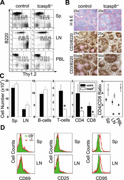
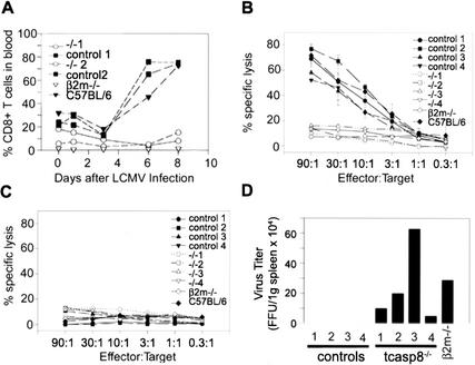
Defects in death receptor-mediated apoptosis have been linked to cancer and autoimmune disease in humans. The in vivo role of caspase 8, a component of this pathway, has eluded analysis in postnatal tissues because of the lack of an appropriate animal model. Targeted disruption of caspase 8 is lethal in utero. We generated mice with a targeted caspase 8 mutation that is restricted to the T-cell lineage. Despite normal thymocyte development in the absence of caspase 8, we observed a marked decrease in the number of peripheral T-cells and impaired T-cell response ex vivo to activation stimuli. caspase 8 ablation protected thymocytes and activated T-cells from CD95 ligand but not anti-CD3-induced apoptosis, or apoptosis activated by agents that are known to act through the mitochondria. caspase 8 mutant mice were unable to mount an immune response to viral infection, indicating that caspase 8 deletion in T-cells leads to immunodeficiency. These findings identify an essential, cell-stage-specific role for caspase 8 in T-cell homeostasis and T-cell-mediated immunity. This is consistent with the recent identification of caspase 8 mutations in human immunodeficiency.


Figures

Figure 1
Figure 1
Generation of conditional caspase 8 mutant mice. (A) Schematic representation of the wild-type caspase 8 locus, the targeting construct, and the caspase 8 mutant alleles casp8fl3-4;neo-tk, casp8fl3-4, and casp8Δ3-4. Exons are indicated as filled boxes and LoxP sites as triangles. B, BamHI site. (B) Four independent clones carrying homologous recombination to the caspase 8 locus were identified by PCR and confirmed by Southern blot with the 5′ probe. (Lane 1) Wild-type ES DNA. (Lanes 25) casp8fl3-4;neo-tk ES clones. (C) casp8fl3-4,neo-tk ES clones were transiently transfected with a CMV-driven construct encoding for Cre recombinase. Clones that have lost the neo-tk cassettes but retained the floxed caspase 8 exons 3 and 4 (casp8fl3-4) were identified by Southern blot with exons 3–4.(Lane 1) Wild-type ES clone. (Lane 2) casp8fl3-4;neo-tk/wt ES clone. (Lane 3) casp8fl3-4/wt ES clone. (Lane 4) casp8Δ3-4/wt ES clone. (D) Heterozygous (casp8fl3-4/wt) mutant littermate mice were identified by Southern blot on tail DNA with a 5′-flanking probe. (Lanes 1,3,4) casp8fl3-4/wt. (Lane 2) Wild type. BamHI restriction fragments (detected by 5′ probe): wild-type fragment = 9.4 kb; casp8fl3-4;neo-tk = 8.7 kb; casp8fl3-4 = 4.5 kb; and casp8Δ3-4 = 9.0 kb.
Figure 2
Figure 2
Deletion of caspase 8 in the T-cell lineage. (A) Southern blot performed on BamHI-digested genomic DNA extracted from tcasp8−/− mice shows a specific and complete deletion of exons 3 and 4 of caspase 8 in thymocytes in comparison to tail extracted DNA. (Lane 1) Wild-type tail DNA. (Lane 2) casp8fl3-4/wt; LckCre tail DNA. (Lane 3) casp8fl3-4/wt; LckCre thymocyte DNA. (Lane 4) tcasp8−/− tail DNA. (Lane 5) tcasp8−/− thymocyte DNA. (B) The casp8Δ3-4 nucleotide sequence contained several termination codons that disrupted the open reading frame of caspase 8 shortly beyond exon 2. (C) Western blot performed on thymocyte cell extracts derived from control and tcasp8−/− mice confirmed the absence of caspase 8 protein in thymocytes. (D) Representative dot blots showing similar thymocyte populations in tcasp8−/− and control mice. (E) Mean total thymocyte numbers in tcasp8−/− and control mice (n = 17, p = 0.601).
Figure 3
Figure 3
Caspase 8 is required for CD95 but not mitochondrial-mediated apoptosis in the T-cell lineage. (A) Sensitivity of thymocytes to DR-mediated apoptosis induced by CD95L and mitochondrial-mediated apoptosis induced by ionizing radiation (γIR) or dexamethasone. Death induced by anti-CD3 and anti-CD28 antibodies and thymocyte death from neglect were also measured. Cell viability was determined 24 h after treatment (n ≥ 3 for each data point). (B) Activated T-cell death was studied in a similar fashion. (C) Thymocytes were treated with 1 μg/mL CD95 ligand for the indicated times. Western blots were performed on lysates with the indicated antibodies. The anti-caspase 8 antibody detects the C-terminal p10 cleavage product of caspase 8 in control but not tcasp8−/− thymocytes. Caspase 3 cleavage was markedly reduced in tcasp8−/− thymocytes treated with CD95L but not in response to γIR. Similarly, loss of full-length Bid was reduced in tcasp8−/− thymocytes in response to CD95L but not to γIR. p53 response to γIR was unaffected by caspase 8 mutation.
Figure 4
Figure 4
Caspase 8 is essential for T-cell homeostasis. (A) Thy1.2+-positive T-cell numbers are decreased in tcasp8−/− compared with control spleen (Sp), lymph nodes (LN), and peripheral blood (PBL). Representative dot blots are shown. (B) Histological examination of tcasp8−/− spleens identifies irregular T- and B-cell follicles characterized by a decreased presence of T-cells (anti-CD3, pink staining) and increased B-cells (anti-B220, brown staining) compared with spleen from control littermates. (C) The mean total number of lymphocytes in the spleens was similar, yet the total number of Thy1.2+ T-cells was significantly decreased in tcasp8−/− spleens. Total CD8+ and to a lesser extent, CD4+ T-cells were reduced in spleen, LN, and PBL from tcasp8−/− mice. The CD4:CD8 ratio was markedly increased in spleen (n = 13), LN (n = 13), and PBL (n = 4). *, p < 0.05. (D) CD69 and CD25 levels were normal on naive caspase 8-deficient peripheral T-cells; however, there was increased expression of CD95 on tcasp8−/− T-cells.
Figure 5
Figure 5
Defective cell growth of tcasp8−/− T-cells. (A) [3H]Thymidine incorporation in tcasp8−/− and control purified T-cells after 48 or 72 h of stimulation with anti-CD3 (2 or 5 μg/mL) with or without costimulation by anti-CD28 (2 μg/mL) or IL-2 (100 U/mL). Representative experiments are shown. (B) [3H]Thymidine incorporation in tcasp8−/− and control purified T-cells after stimulation with PMA and ionomycin (concentrations in nanograms per milliliter). Representative experiments are shown. (C) [3H]Thymidine incorporation in B-cells purified from tcasp8−/− and control mice after 48 or 72 h of activation with various stimuli. (D) The proportion of T-cells in G0/G1, S, and G2/M phases were similar in control and tcasp8−/− backgrounds after anti-CD3 and anti-CD28 stimulation. The larger G1 subpopulation in the cycling tcasp8−/− T-cells at 48 and 72 h after stimulation indicates decreased survival of stimulated tcasp8−/− T-cells. G0/G1, S, and G2/M are calculated from the total number of cycling T-cells. Sub-G1 values represent the percentage of the total number of all cells cycling or dead.
Figure 6
Figure 6
Activation of tcasp8−/− T-cells. A. The profile of tyrosine-phosphorylated proteins was similar in tcasp8−/− and control T-cells activated with anti-CD3 antibody for 5 min. (B) NF-κB activation analyzed by gel shift was found to be similar in control and tcasp8−/− T-cells in response to various stimuli. (C) Baseline levels and phosphorylation of p42/44-MAPK (Erk1/2) following anti-CD3 activation of purified T-cells was similar in control and tcasp8−/− T-cells. (D) IL-2 concentration in the supernatant of purified T-cells after 48 and 72 h of stimulation with anti-CD3 and anti-CD28 antibodies. A representative experiment is shown. (E) The expression of CD95 and the activation markers CD44, CD69, and CD25 (IL-2Rα) were similarly up-regulated on tcasp8−/− and control T-cells at 48 and 72 h after anti-CD3 and anti-CD28 stimulation.
Figure 7
Figure 7
Defective T-cell response of tcasp8−/− mice to LCMV infection. (A) The expansion of CD8+ T-cells in peripheral blood was monitored by flow cytometry using specific antibodies for Thy1.2, CD4, and CD8 on days 0, 1, 3, 6, and 8. (B,C) The cytotoxic response of splenocytes was assessed 8 d after LCMV infection. EL4 cells were prepulsed with p33 (a specific LCMV peptide) or AV (a control peptide) and were used as target cells. (D) Viral titer was assessed from the spleens of mice 8 d after LCMV infection.

References

    1. Alam A, Cohen LY, Aouad S, Sekaly RP. Early activation of caspases during T lymphocyte stimulation results in selective substrate cleavage in nonapoptotic cells. J Exp Med. 1999;190:1879–1890. - PMC - PubMed
    1. Ashkenazi A. Targeting death and decoy receptors of the tumour-necrosis factor superfamily. Nat Rev Cancer. 2002;2:420–430. - PubMed
    1. Bergeron L, Perez GI, Macdonald G, Shi L, Sun Y, Jurisicova A, Varmuza S, Latham KE, Flaws JA, Salter JC, et al. Defects in regulation of apoptosis in caspase-2-deficient mice. Genes & Dev. 1998;12:1304–1314. - PMC - PubMed
    1. Berry DM, Benn SJ, Cheng AM, McGlade CJ. Caspase-dependent cleavage of the hematopoietic specific adaptor protein Gads alters signalling from the T cell receptor. Oncogene. 2001;20:1203–1211. - PubMed
    1. Blattman JN, Antia R, Sourdive DJ, Wang X, Kaech SM, Murali-Krishna K, Altman JD, Ahmed R. Estimating the precursor frequency of naive antigen-specific CD8 T cells. J Exp Med. 2002;195:657–664. - PMC - PubMed

Publication types