. 2003 Apr;71(4):1961-71.
doi: 10.1128/IAI.71.4.1961-1971.2003.

Signaling through the T1/ST2 molecule is not necessary for Th2 differentiation but is important for the regulation of type 1 responses in nonhealing Leishmania major infection

Affiliations

Signaling through the T1/ST2 molecule is not necessary for Th2 differentiation but is important for the regulation of type 1 responses in nonhealing Leishmania major infection

P Kropf et al. Infect Immun. 2003 Apr.

Abstract

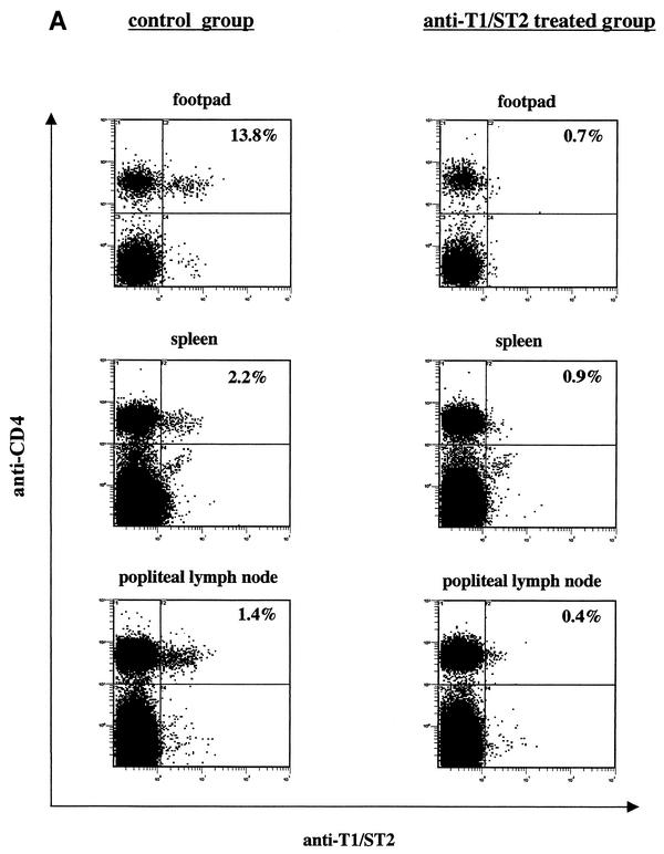
T1/ST2 is a stable cell surface marker selectively expressed on type 2 T helper (Th2) effector cells. Since nonhealing Leishmania major infections in susceptible BALB/c mice have been ascribed to a polarized Th2 response, we used an anti-T1/ST2 monoclonal antibody (MAb) or a T1-Fc fusion protein to investigate the role of CD4+ T1/ST2(+) Th2 cells in experimental leishmaniasis. We show that interfering with T1/ST2 signaling had no effect on lesion development or parasite replication; however, it induced a significantly higher type 1 response and an enhanced capacity of CD4+ T cells to respond to interleukin 12 (IL-12). Surprisingly, even in the presence of an elevated Th1 response, the production of antigen-specific type 2 cytokines was not altered in the group of mice treated with the anti-T1/ST2 MAb or the T1-Fc fusion protein. To characterize further this Th2 response, we assessed the cytokine profile of CD4+ T cells and found that interfering with T1/ST2 signaling did not alter the cytokine profile of CD4+ T1/ST2(+) T cells. These results show that T1/ST2 signaling is not necessary for the differentiation of naive CD4+ T cells into antigen-specific CD4+ T1/ST2(+) Th2 cells. In addition to CD4+ T1/ST2(+) T cells, we detected another subpopulation of CD4+ Th2 cells, negative for the expression of T1/ST2, that could differentiate in vivo in response to L. major infection. Taken together, our results suggest that CD4+ T1/ST2(+) Th2 cells but not CD4+ T1/ST2(-) Th2 cells can downregulate the Th1 response during the course of a nonhealing L. major infection through a mechanism that is independent of IL-4 or IL-10.


Figures

FIG. 1.
FIG. 1.
Lesion development and parasite load. (A) Lesion development and parasite load in L. major-infected BALB/c mice treated with anti-T1/ST2 MAb. Groups of L. major-infected BALB/c mice (n = 4) were treated twice a week throughout the course of infection with 80 μg of anti-T1/ST2 MAb or 80 μg of rat IgG injected i.p., and one group was used as a control. Lesion development was monitored by measuring the increase in footpad thickness. At 5 weeks postinfection, the numbers of viable parasites in the lesions of individual mice were determined by an LDA. (B) Parasite load in L. major-infected BALB/c mice treated with T1-Fc fusion protein. Groups of L. major-infected BALB/c mice (n = 4) were treated twice a week throughout the course of infection with 400 μg of T1-Fc fusion protein injected subcutaneously in the footpads, and one group was used as a control. At 5 weeks postinfection, the numbers of viable parasites in the lesions of individual mice were determined by an LDA. Each symbol represents one mouse, and the horizontal lines represent the averages for four mice per group. Data show the results of one representative experiment out of three independent experiments. (C) Biological activity of T1-Fc fusion protein. BMM (5 × 10⁵/ml) were stimulated with 10 ng of LPS in the presence or in the absence of 500 μg of T1-Fc fusion protein. After 24 h, supernatants were harvested and tested for the presence of IL-6 by an ELISA. Error bars in panels A and C indicate standard errors of the means.
FIG. 2.
FIG. 2.
Anti-T1/ST2 MAb binds to but does not lyse CD4+ T1/ST2+ T cells in vivo. One group of L. major-infected BALB/c mice (n = 4) was treated twice a week throughout the course of infection with 80 μg of anti-T1/ST2 MAb, and one group was used as a control. At 5 weeks postinfection, the expression of T1/ST2 on CD4+ T cells was determined directly ex vivo in the footpads, spleens, and draining lymph nodes (A) or after in vitro restimulation of spleen and lymph node cells with L. major parasites (B). Data show the results for one representative mouse per group, and similar results were obtained in three independent experiments.
FIG. 2.
FIG. 2.
Anti-T1/ST2 MAb binds to but does not lyse CD4+ T1/ST2+ T cells in vivo. One group of L. major-infected BALB/c mice (n = 4) was treated twice a week throughout the course of infection with 80 μg of anti-T1/ST2 MAb, and one group was used as a control. At 5 weeks postinfection, the expression of T1/ST2 on CD4+ T cells was determined directly ex vivo in the footpads, spleens, and draining lymph nodes (A) or after in vitro restimulation of spleen and lymph node cells with L. major parasites (B). Data show the results for one representative mouse per group, and similar results were obtained in three independent experiments.
FIG. 3.
FIG. 3.
Treatment of L. major-infected BALB/c mice with anti-T1/ST2 MAb does not alter the production of Th2 cytokines. One group of L. major-infected BALB/c mice (n = 4) was treated twice a week throughout the course of infection with 80 μg of anti-T1/ST2 MAb, and one group was used as a control. At 5 weeks postinfection, lymph node cells were restimulated with L. major parasites. After 3 days, supernatants were harvested and tested for Th2 cytokine contents by an ELISA. Each symbol represents one mouse, and the horizontal lines represent the averages for four mice per group. Data show the results of one representative experiment out of three independent experiments.
FIG. 4.
FIG. 4.
Treatment of L. major-infected BALB/c with anti-T1/ST2 MAb or T1-Fc fusion protein induces a strong type 1 response. (A) One group of L. major-infected BALB/c mice (n = 4) was treated twice a week throughout the course of infection with 80 μg of anti-T1/ST2 MAb, and one group was used as a control. At 5 weeks postinfection, lymph node cells were restimulated with L. major parasites. After 3 days, supernatants were harvested and tested for IFN-γ content by an ELISA. Each symbol represents one mouse, and the horizontal lines represent the averages for four mice per group. Data show the results of one representative experiment out of three independent experiments. (B) An experiment similar to that shown in panel A was performed with 80 or 400 μg of T1-Fc fusion protein. The error bar shows the standard error of the mean.
FIG. 5.
FIG. 5.
CD4+ T cells from mice treated with anti-T1/ST2 MAb or T1-Fc fusion protein have an enhanced capacity to produce IFN-γ in response to IL-12. (A) One group of L. major-infected BALB/c mice (n = 4) was treated twice a week throughout the course of infection with 80 μg of anti-T1/ST2 MAb, and one group was used as a control. At 5 weeks postinfection, CD4+ T cells were purified from the lymphoid organs and restimulated in the presence of L. major-infected APCs and IL-12 (10 U/ml); the levels of IFN-γ produced in response to IL-12 were determined by an ELISA. As controls, CD4+ T cells were stimulated with APCs and antigen in the absence of IL-12. The levels of IFN-γ detected were 4.9 U/ml for the control group and 7.4 U/ml for the group treated with anti-T1/ST2 MAb, and no IFN-γ was detectable in the absence of IL-12 and antigen (data not shown). Data show the results of one representative experiment out of three independent experiments. (B) An experiment similar to that shown in panel A was performed with 80 or 400 μg of T1-Fc fusion protein. As controls, CD4+ T cells were stimulated with APCs and antigen in the absence of IL-12. The levels of IFN-γ detected were 9 U/ml for the group treated with 80 μg of T1-Fc fusion protein and 16 U/ml for the group treated with 400 μg of T1-Fc fusion protein, no IFN-γ was detectable in the control group, and no IFN-γ was detectable in the absence of IL-12 and antigen (data not shown). Error bars in both panels indicate standard errors of the means.
FIG. 6.
FIG. 6.
Interfering with T1/ST2 signaling does not influence the frequencies of IL-4- and IL-10-producing CD4+ T cells. One group of L. major-infected BALB/c mice (n = 4) was treated twice a week throughout the course of infection with 80 μg of T1-Fc fusion protein, and one group was used as a control. At 5 weeks postinfection, lymph node cells were restimulated with L. major parasites. After 6 days, viable cells were purified with a Ficoll gradient and stimulated with phorbol 12-myristate 13-acetate (50 ng) and ionomycin (500 ng) for 4 h; brefeldin A (10 μg) was included during the last 2 h. Cells were labeled with anti-CD4, anti-T1/ST2, anti-IL-4, or anti-IL-10. The expression of IL-4 and IL-10 in CD4+ T1/ST2+ T cells (A, C, E, and G) and CD4+ T1/ST2 T cells (B, D, F, and H) was determined by flow cytometry. An isotype control (IgG1-PE) was used at the same concentration as the antibodies, and 1% of the cells stained positive. Similar results were obtained in two independent experiments.

References

    1. Coyle, A. J., C. Lloyd, T. Nguyen, C. Erikson, L. Wang, P. Ottonson, P. Persson, T. Delaney, S. Lehar, S. Lin, L. Poisson, C. Meisel, T. Kamradt, T. Bjerke, D. Levinson, and J.-C. Gutierrez-Ramos. 1999. Crucial role of IL-1 receptor family member T1/ST2 in T helper cell type 2-mediated lung mucosal immune response. J. Exp. Med. 190:895-902. - PMC - PubMed
    1. Etges, B., and I. Müller. 1998. Progressive disease or protective immunity to Leishmania major infection: the result of a network of stimulatory and inhibitory interactions. J. Mol. Med. 76:372-390. - PubMed
    1. Himmelrich, H., P. Launois, I. Maillard, T. Biedermann, F. Tacchini-Cottier, R. M. Locksley, M. Röcken, and J. A. Louis. 2000. In BALB/c mice, IL-4 production during the initial phase of infection with L. major is necessary and sufficient to instruct Th2 cell development resulting in progressive disease. J. Immunol. 164:4819-4825. - PubMed
    1. Hondowicz, B., A. Y. Park, M. M. Elloso, and P. Scott. 2000. Maintenance of IL-12-responsive CD4+ T cells during a Th2 response in L. major-infected mice. Eur. J. Immunol. 30:2007-2014. - PubMed
    1. Hondowicz, B. D., T. M. Scharton-Kersten, D. Jones, and P. Scott. 1997. Leishmania major-infected C3H mice treated with anti-IL-12 mAb develop but do not maintain a Th2 response. J. Immunol. 159:5024-5031. - PubMed

Publication types

MeSH terms