. 2003 Apr;71(4):1995-2001.
doi: 10.1128/IAI.71.4.1995-2001.2003.

Anaplasma phagocytophilum reduces neutrophil apoptosis in vivo

Affiliations
Free PMC article

Anaplasma phagocytophilum reduces neutrophil apoptosis in vivo

Helena Scaife et al. Infect Immun. 2003 Apr.
Free PMC article

Abstract

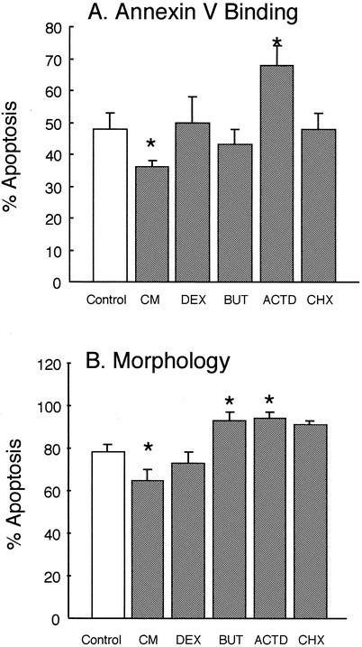
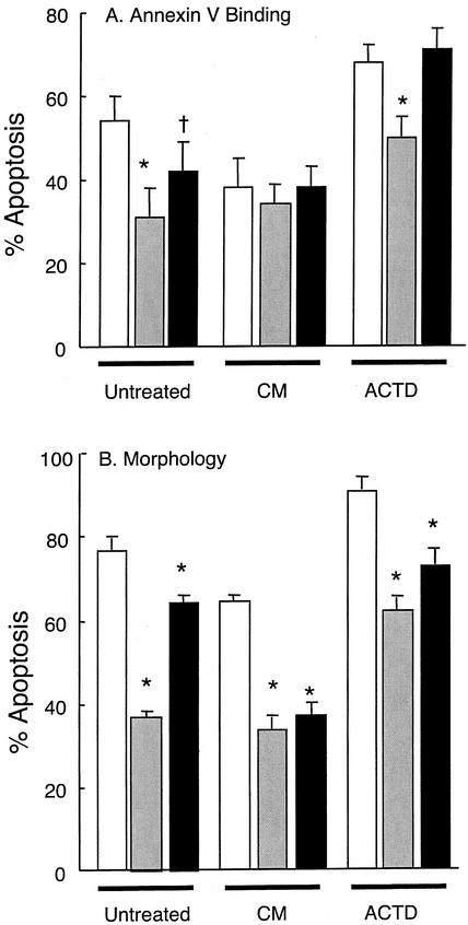
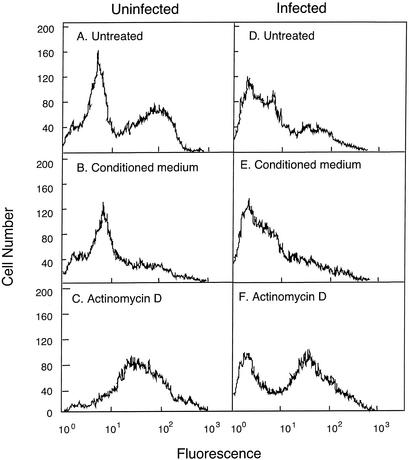
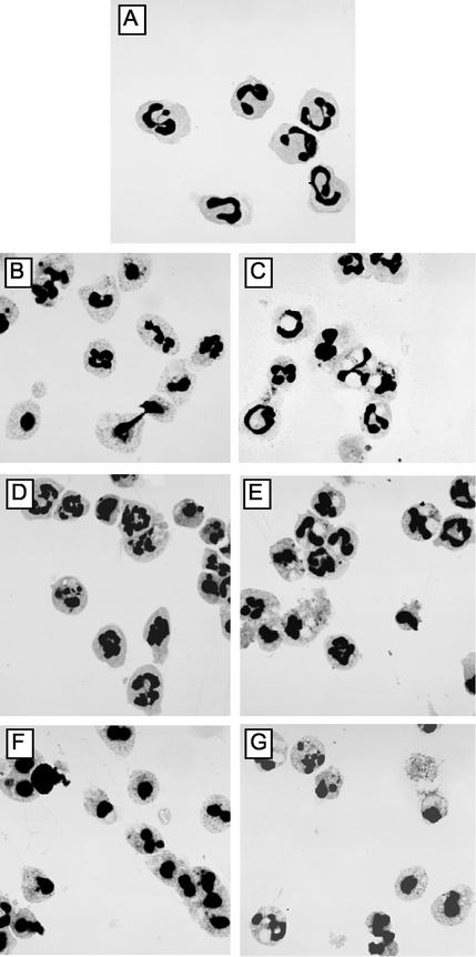
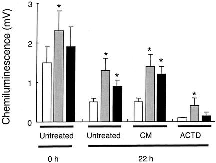
Ovine neutrophils spontaneously underwent apoptosis during culture in vitro, as assessed by morphological changes and exposure of annexin V binding sites on their cell surfaces. The addition of conditioned medium from concanavalin A-treated ovine peripheral blood mononuclear cells (PBMC) could partially protect against this progression into apoptosis, but dexamethasone and sodium butyrate could not. Actinomycin D accelerated the rate at which ovine neutrophils underwent apoptosis. Neutrophils isolated from sheep experimentally infected with Anaplasma phagocytophilum showed significantly delayed apoptosis during culture ex vivo, and the addition of conditioned medium from PBMC to these cells could not delay apoptosis above the protective effects observed after in vivo infection. The ability of neutrophils from A. phagocytophilum-infected sheep to activate a respiratory burst was increased compared to the activity measured in neutrophils from uninfected sheep, but chemotaxis was decreased in neutrophils from infected sheep. These data are the first demonstration that in vivo infection with A. phagocytophilum results in changes in rates of apoptosis of infected immune cells. This may help explain how these bacteria replicate in these normally short-lived cells.


Figures

FIG. 1.
FIG. 1.
Effects of exogenous agents on apoptosis of ovine neutrophils. Purified ovine neutrophils were incubated in RPMI 1640 medium supplemented with 10% fetal bovine serum and 2 mM l-glutamine at 2.5 × 10⁶ cells/ml. They were then incubated for 22 h at 37°C as follows: Control, no additions; CM, conditioned medium obtained from concanavalin A-stimulated PBMCs, as described in Materials and Methods; DEX, dexamethasone at 1 μM; BUT, sodium butyrate at 0.4 mM; ACTD, actinomycin D at 1 μM; CHX, cycloheximide at 10 μg/ml. After culture, apoptosis was assessed by binding to FITC-annexin V and flow cytometry (A) and by morphological examination of cytospins (B). Immediately after isolation, neutrophils were 100% nonapoptotic, as assessed by these criteria. Values shown are means ± the SD (n = 6). Values indicated by asterisks are significantly different from the control values (P < 0.05).
FIG. 2.
FIG. 2.
Effects of A. phagocytophilum infection on neutrophil apoptosis. Neutrophils were isolated from the venous blood of sheep prior to infection (□) and on the second (░⃞) and fourth (▪) days of bacteremia. Purified neutrophils were then incubated in RPMI 1640 medium as described in the legend to Fig. 1. Cultures contained no further additions (Untreated), PBMC conditioned medium (CM), or actinomycin D (ACTD). After culture with gentle agitation at 37°C for 22 h, apoptosis was determined by FITC-annexin V binding (A) and morphology (B). Values shown are means ± the SD (n = 6). Values statistically different to the preinfection values are indicated by an asterisk (P = 0.01) or a dagger (P = 0.05).
FIG. 3.
FIG. 3.
Changes in annexin V binding to ovine neutrophils. Neutrophils were isolated from ovine blood prior to infection (Uninfected) and on the second day of bacteremia (Infected) after experimental infection of the sheep with A. phagocytophilum. The purified neutrophils were then incubated in the absence (Untreated, A and D) or the presence of PBMC conditioned medium (B and E) or Actinomycin D (C and D) for 22 h at 37°C with gentle agitation, as described in the legend to Fig. 1. They were then labeled with FITC-annexin V and examined by flow cytometry. The data shown are from an individual sheep before and after infection. Similar results were obtained for five other sheep.
FIG. 4.
FIG. 4.
Changes in neutrophil morphology during culture. Cytospins were prepared from purified neutrophil suspensions from uninfected (A, B, D, and F) and from A. phagocytophilum-infected sheep at the second day of bacteremia (C, E, and G). Neutrophils were incubated as described in the legend to Fig. 1. (A) Neutrophil morphology immediately after isolation, with cells showing no morphological features of apoptosis. Neutrophils incubated for 22 h at 37°C in RPMI 1640 medium supplemented with 10% fetal bovine serum and l-glutamine only (B and C), while panels D and E also contained PBMC conditioned medium and panels F and G were supplemented with actinomycin D. The results shown are typical of those obtained from six different sheep.
FIG. 5.
FIG. 5.
Changes in NADPH oxidase activity of neutrophils isolated from uninfected and A. phagocytophilum-infected sheep. Neutrophils were isolated from the venous blood of sheep prior to infection (□) and on the second (░⃞) and fourth (▪) days of bacteremia. Purified neutrophils were then incubated in RMPI 1640 medium as described in the legend to Fig. 1. Cultures contained no further additions (Untreated), PBMC conditioned medium (CM), or actinomycin D (ACTD). The activity of the NADPH oxidase was then measured after incubation for 0 and 22 h by luminol chemiluminescence after the addition of 0.1 μg of PMA/ml. Values shown are means ± the SD (n = 6), and asterisk indicates values significantly different from the value measured in neutrophils from uninfected sheep (P < 0.02).

Similar articles

Cited by

References

    1. Aga, E., D. M. Katschinski, G. Van Zandbergen, H. Laufs, B. Hansen, K. Muller, W. Solbach, and T. Laskay. 2002. Inhibition of the spontaneous apoptosis of neutrophil granulocytes by the intracellular parasite Leishmania major. J. Immunol. 169:898-905. - PubMed
    1. Akgul, C., D. A. Moulding, and S. W. Edwards. 2001. Molecular control of neutrophil apoptosis. FEBS Lett. 487:318-322. - PubMed
    1. Bakken, J. S., J. S. Dumler, S. M. Chen, M. R. Eckman, L. L. Van Etta, and D. H. Walker. 1994. Human granulocytic ehrlichiosis in the upper midwest United States: a new emerging species? JAMA 272:212-218. - PubMed
    1. Banerjee, R., J. Anguita, D. Roos, and E. Fikrig. 2000. Cutting edge: infection by the agent of human granulocytic ehrlichiosis prevents the respiratory burst by downregulating gp91phox1. J. Immunol. 164:3946-3949. - PubMed
    1. Bedner, E., P. Burfeind, T.-C Hseih, J. M. Wu, M. E. Aguero-Rosenfeld, M. R. Melamed, H. W. Horowitz, G. P. Wormser, and Z. Darzynkiewicz. 1998. Cell cycle effects and induction of apoptosis caused by infection of HL-60 cells with human granulocytic ehrlichiosis pathogen measured by flow and laser scanning cytometry. Cytometry 33:47-55. - PubMed

Publication types

LinkOut - more resources