. 2003 Apr;71(4):2065-70.
doi: 10.1128/IAI.71.4.2065-2070.2003.

Exposure of immunocompetent adult mice to Pneumocystis carinii f. sp. muris by cohousing: growth of P. carinii f. sp. muris and host immune response

Affiliations

Exposure of immunocompetent adult mice to Pneumocystis carinii f. sp. muris by cohousing: growth of P. carinii f. sp. muris and host immune response

Chun Li An et al. Infect Immun. 2003 Apr.

Abstract

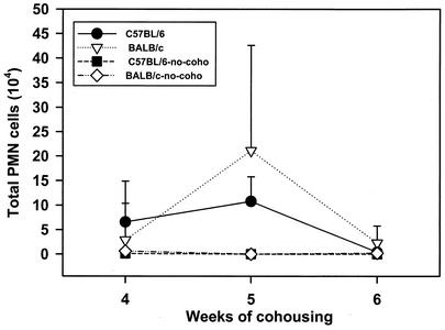
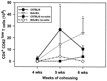
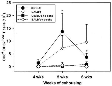
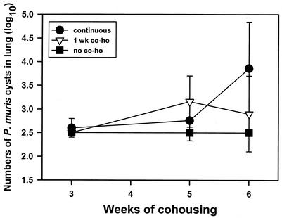
There has been emerging evidence that immunocompetent hosts can harbor Pneumocystis in their lungs. The purpose of this study was to determine the kinetics of Pneumocystis carinii f. sp. muris infection in adult immunocompetent mice and the host immune response to the organisms. To accomplish this, we exposed adult immunocompetent mice to SCID mice infected with P. carinii f. sp. muris by cohousing. We found that P. carinii f. sp. muris was detectable in the lungs of cohoused immunocompetent mice by PCR by 3 weeks after the beginning of cohousing. At about 4 weeks of cohousing, P. carinii f. sp. muris was readily detectable in the lungs of mice by microscopic techniques. Also at this time, P. carinii f. sp. muris-specific immunoglobulin G was found in the sera of the mice, and CD62(low) CD4- and CD8-positve T cells accumulated in the lungs. Shortly after this immune response, the P. carinii f. sp. muris organisms were cleared from the lungs. Adult mice cohoused for only 1 week also contained P. carinii f. sp. muris cysts detectable by silver staining at 5 and 6 weeks after the beginning of cohousing. We also found that the P. carinii f. sp. muris organisms grew to greater numbers in the lungs of BALB/c mice than in those of C57BL6 mice. This indicates that immunocompetent hosts develop a mild infection with P. carinii f. sp. muris which resolves in 5 to 6 weeks when there is a detectable immune response to the organism. Once an acquired immune response was initiated, the P. carinii f. sp. muris organisms were quickly eliminated without clinical signs of disease.


Figures

FIG. 1.
FIG. 1.
Numbers of P. carinii f. sp. muris cysts (log10) in lungs of C57BL/6 and BALB/c mice with and without (no-coho) cohousing with P. carinii f. sp. muris-infected SCID mice. Mice were cohoused with P. carinii f. sp. muris-infected SCID mice for 4, 5, or 6 weeks; the mice were then killed, and the numbers of P. carinii f. sp. muris cysts in their lungs were determined by silver staining of lung homogenates. Other mice were not cohoused as a control. The minimal level of detection of cysts was 102.5. Data points are the average number of cysts ± standard deviation of the mean at each time point (n = 5). ∗, P < 0.05 for comparison of cohoused and control mice, as calculated by one-way ANOVA.
FIG. 2.
FIG. 2.
Numbers of P. carinii f. sp. muris organisms (log10) in lungs of C57BL/6 and BALB/c mice with and without cohousing with P. carinii f. sp. muris-infected SCID mice. Mice were cohoused with P. carinii f. sp. muris-infected SCID mice for 15, 20, 25, 30, 35, 40, or 45 days; the mice were then killed, and the numbers of P. carinii f. sp. muris organisms in their lungs were determined by immunostaining of lung homogenates. The minimal level of detection was 103.1 organisms. The numbers of P. carinii f. sp. muris organisms are the average number of organisms ± standard deviation of the mean at each time point (n = 5). ∗, P < 0.05 for comparison of results at day 15 and those at days 30 and 35, as calculated by one-way ANOVA.
FIG. 3.
FIG. 3.
Numbers of polymorphonuclear neutrophils (PMN) in lung lavage fluids of C57BL/6 and BALB/c mice after cohousing. Mice were cohoused with P. carinii f. sp. muris-infected SCID mice for 4, 5, or 6 weeks; the mice were then killed, and the numbers of PMN in their lung lavage fluids were determined. Other mice (no-coho) were not cohoused as control. Data points are the average numbers of PMN ± standard deviation of the mean at each time point (n = 5).
FIG. 4.
FIG. 4.
Numbers of CD4+ CD62low T cells in lung lavage fluids of C57BL/6 and BALB/c mice after cohousing. Mice were cohoused with P. carinii f. sp. muris-infected SCID mice for 4, 5, or 6 weeks; the mice were then killed, and the numbers of CD4+ CD62low T cells in their lung lavage fluids were determined. Other mice (no-coho) were not cohoused as a control. Data points are the average numbers of CD4+ CD62low T cells ± standard deviation of the mean at each time point (n = 5). ∗, P < 0.05 for comparison of cohoused and control mice, as calculated by one-way ANOVA.
FIG. 5.
FIG. 5.
Numbers of CD8+ CD62low T cells in lung lavage fluids of C57BL/6 and BALB/c mice after cohousing. Mice were cohoused with P. carinii f. sp. muris-infected SCID mice for 4, 5, or 6 weeks; the mice were then killed, and the numbers of CD8+ CD62low T cells in their lung lavage fluids were determined. Other mice (no-coho) were not cohoused as a control. Data points are the average numbers of CD8+ CD62low T cells ± standard deviation of the mean at each time point (n = 5). ∗, P < 0.05 for comparison of cohoused and control mice, as calculated by one-way ANOVA.
FIG. 6.
FIG. 6.
P. carinii f. sp. muris-specific IgG in lung lavage fluid of C57BL/6 and BALB/c mice after cohousing. Mice were cohoused with P. carinii f. sp. muris-infected SCID mice for 4, 5, or 6 weeks; the mice were then killed, and the levels of P. carinii f. sp. muris-specific IgG in their lung lavage fluids were determined by ELISA. Other mice (no-coho) were not cohoused as a control. Data points are the average optical densities ± standard deviation of the mean at each time point (n = 5).
FIG. 7.
FIG. 7.
Numbers of P. carinii f. sp. muris cysts (log10) in lungs of CB-17 mice with 1 week of cohousing (1 wk co-ho) or continuous cohousing with P. carinii f. sp. muris-infected SCID mice. Mice were cohoused with P. carinii f. sp. muris-infected SCID mice for 3, 4, 5, or 6 weeks; the mice were then killed, and the numbers of P. carinii f. sp. muris cysts in their lungs were determined by silver staining of lung homogenates. Other mice (no co-ho) were not cohoused as a control. The minimal level of detection of cysts was 102.5. Data points are the average numbers of cysts ± standard deviation of the mean at each time point (n = 5).

References

    1. Baughman, R. P., S. S. Strohofer, B. A. Clinton, A. D. Nickol, and P. T. Frame. 1989. The use of an indirect fluorescent antibody test for detecting Pneumocystis carinii. Arch. Pathol. Lab. Med. 113:1062-1065. - PubMed
    1. Beck, J. M., M. L. Warnock, J. L. Curtis, M. J. Sniezek, S. M. Arraj-Peffer, H. B. Kaltreider, and J. E. Shellito. 1991. Inflammatory responses to Pneumocystis carinii in mice selectively depleted of helper T lymphocytes. Am. J. Respir. Cell Mol. Biol. 5:186-197. - PubMed
    1. Bille-Hansen, V., S. E. Jorsal, S. A. Henriksen, and O. P. Settnes. 1990. Pneumocystis carinii pneumonia in Danish piglets. Vet. Rec. 127:407-408. - PubMed
    1. Chen, W., F. Gigliotti, and A. G. Harmsen. 1993. Latency is not an inevitable outcome of infection with Pneumocystis carinii. Infect. Immun. 61:5406-5409. - PMC - PubMed
    1. Chen, W., E. A. Havell, F. Gigliotti, and A. G. Harmsen. 1993. Interleukin-6 production in a murine model of Pneumocystis carinii pneumonia: relation to resistance and inflammatory response. Infect. Immun. 61:97-102. - PMC - PubMed

Publication types