. 2003 Apr;71(4):2153-62.
doi: 10.1128/IAI.71.4.2153-2162.2003.

Involvement of myeloid dendritic cells in the development of gastric secondary lymphoid follicles in Helicobacter pylori-infected neonatally thymectomized BALB/c mice

Affiliations

Involvement of myeloid dendritic cells in the development of gastric secondary lymphoid follicles in Helicobacter pylori-infected neonatally thymectomized BALB/c mice

Toshiki Nishi et al. Infect Immun. 2003 Apr.

Abstract

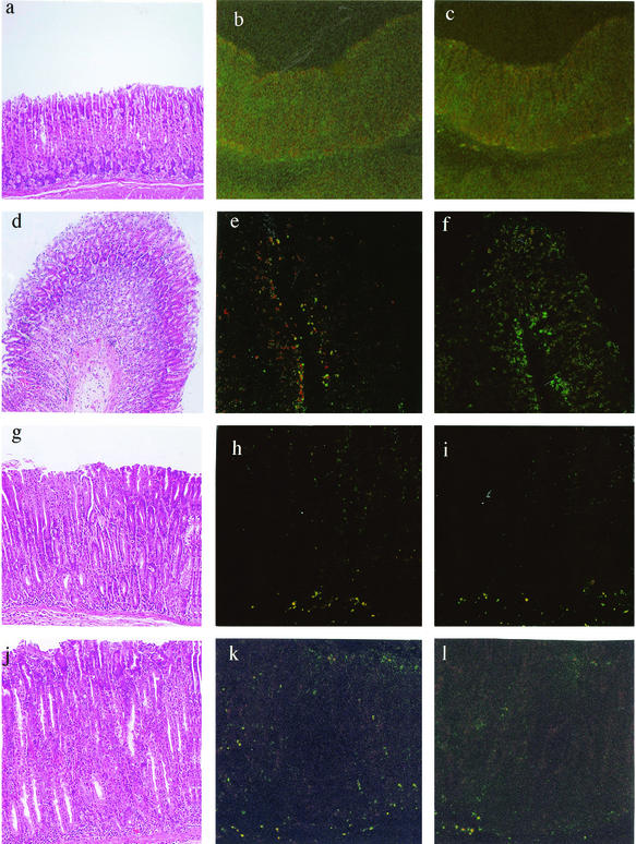
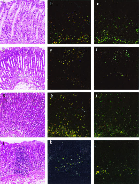
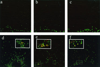
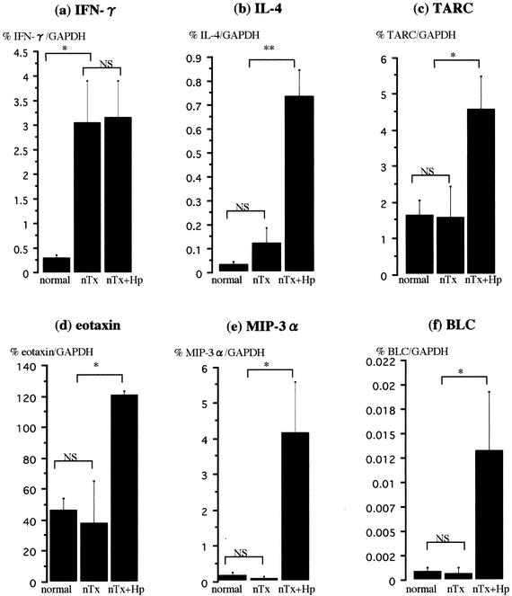
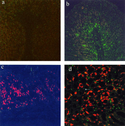
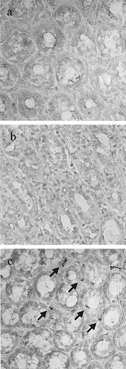
We previously described an animal model of Helicobacter pylori-induced follicular gastritis in neonatally thymectomized (nTx) mice. However, it is still not clear whether antigen-presenting dendritic cells (DCs) in the stomach have a role in the development of secondary follicles in H. pylori-infected nTx mice. We investigated the distribution of DC subsets using this model and examined their roles. To identify lymphoid and myeloid DCs, sections were stained with anti-CD11c (pan-DC marker) in combination with anti-CD8alpha (lymphoid DC marker) or anti-CD11b (myeloid DC marker) and were examined with a confocal microscope. Expression of macrophage inflammatory protein 3alpha (MIP-3alpha), which chemoattracts immature DCs, was analyzed by real-time PCR and immunohistochemistry. Follicular dendritic cells (FDCs) were stained with anti-SKY28 antibodies. In noninfected nTx mice, a few myeloid and lymphoid DCs were observed in the bottom portion of the lamina propria, whereas in H. pylori-infected nTx mice, there was an increased influx of myeloid DCs throughout the lamina propria. FDC staining was also observed in the stomachs of members of the infected group. MIP-3alpha gene expression was upregulated in the infected nTx group, and the immunohistochemistry analysis revealed MIP-3alpha-positive epithelial cells. These data suggest that H. pylori infection upregulates MIP-3alpha gene expression in gastric epithelial cells and induces an influx of myeloid DCs in the lamina propria of the gastric mucosa in nTx mice. Myeloid DCs and FDCs might contribute to the development of gastric secondary lymphoid follicles in H. pylori-infected nTx mice.


Figures

FIG. 1.
FIG. 1.
Confocal microscopic analysis of myeloid and lymphoid DCs in the stomach and hematoxylin and eosin staining of non-nTx (normal) and nTx mice. Two-color staining was performed with 6-μm frozen stomach sections by using Abs against CD11c (green) in combination with anti-CD11b (red) (b, e, h, and k) or anti-CD8α (red) (c, f, i, and l). Sections were prepared from 6-week-old noninfected normal mice (a to c), 6-week-old noninfected nTx mice (d to f), 14-week-old noninfected nTx mice (g to i), and 18-week-old noninfected nTx mice (j to l). DCs positive for either CD11b or CD8α were stained yellow. Sections of the same animals were stained with hematoxylin and eosin (a, d, g, and j) to visualize stomach morphology, and the presence of gastritis in nTx mice was confirmed. There was no apparent gastritis (a) and no DC staining (b and c) in normal mice. The histologic findings for the stomachs of nTx mice were typical of AIG, with a loss of parietal cells, proliferation of pit cells, and moderate to severe lymphocyte infiltration (d, g, and j). In 6-week-old nTx mice, staining of a few myeloid DCs was observed at the bottom of the lamina propria (e), whereas there was no lymphoid DC staining (f). In 14- and 18-week-old nTx mice, some staining of both myeloid and lymphoid DCs was observed in the bottom portion of the lamina propria.
FIG. 2.
FIG. 2.
Confocal microscopic analysis of myeloid and lymphoid DCs in the stomach and hematoxylin and eosin staining of H. pylori-infected nTx mice. The histologic findings for the stomachs from H. pylori-infected nTx mice were mild to severe gastritis (a, d, and g [1, 4, and 8 weeks after infection, respectively]). At 12 weeks after infection (18-week-old mice), there was follicle formation (j). Staining of lymphoid DCs (CD11c and CD8α double positive; yellow) was observed in the bottom portion of the lamina propria and submucosal area throughout the experiment (c, f, i, and l). In contrast, there were myeloid DCs (CD11c and CD11b double positive; yellow) throughout the lamina propria, and the number increased linearly during the observation period (b, e, and h). In H. pylori-infected nTx mice at 12 weeks after infection (18 weeks old), myeloid DCs were induced around the follicle (k).
FIG. 3.
FIG. 3.
Confocal microscopic analysis of activated DCs in the stomachs of H. pylori-infected and noninfected nTx mice (12 weeks after infection, 18 weeks old). Two-color staining was performed with sections by using Abs against CD11c (green) in combination with anti-IAb (red) (a and d), anti-CD80 (red) (b and e), or anti-CD86 (red) (c and f). In the stomachs of noninfected nTx mice, there was no staining of double-positive cells (a, b, and c). In contrast, there were a few double-positive cells in the bottom portions of the lamina propria in the stomachs of H. pylori-infected nTx mice (yellow) (d, e, and f) (the small boxes were magnified to produce the large boxes).
FIG. 4.
FIG. 4.
Profiles of cytokine and chemokine mRNA expression in the gastric mucosa of non-H. pylori-infected normal mice (non-nTx, 14 weeks old), non-H. pylori-infected nTx mice (14 weeks old), and H. pylori-infected nTx mice (8 weeks after infection, 14 weeks old), as determined by real-time PCR. Ten mice from each group were analyzed. The data are expressed as the means ± standard errors for the number of copies of the mRNA per GAPDH gene copy. One asterisk and two asterisks indicate that the data were significantly different at P values of <0.05 and <0.01, respectively. NS, not significant; Hp, H. pylori infected.
FIG. 5.
FIG. 5.
Confocal microscopic analysis of DCs and MIP-3α staining in the gastric mucosa of non-H. pylori-infected normal mice (non-nTx) (a), H. pylori-infected normal mice (non-nTx, 14 weeks old) (b), non-H. pylori-infected nTx mice (14 weeks old) (c), and H. pylori-infected nTx mice (8 weeks after infection, 14 weeks old) (d). Two-color staining of the sections was performed with Ab against CD11c (green) in combination with anti-MIP-3α (red).
FIG. 6.
FIG. 6.
Immunohistochemical staining of FDCs in the gastric mucosa of nTx mice (original magnification, ×400). Gastric tissues were incubated with isotype-matched control goat immunoglobulin G (a) and anti-mouse SKY28 Ab (b and c). Positive cells (arrows) were observed in the stomachs of H. pylori-infected nTx mice (8 weeks after infection, 14 weeks old) (c), whereas there were no positive cells in the stomachs of noninfected nTx mice (14 weeks old) (b).

Similar articles

Cited by

References

    1. De Togni, P., J. Goellner, N. H. Ruddle, P. R. Streeter, A. Fick, S. Mariathasan, S. C. Smith, R. Carlson, L. P. Shornick, J. Strauss-Schoenberger, et al. 1994. Abnormal development of peripheral lymphoid organs in mice deficient in lymphotoxin. Science 264:703-707. - PubMed
    1. Dieu-Nosjean, M. C., A. Vicari, S. Lebecque, and C. Caux. 1999. Regulation of dendritic cell trafficking: a process that involves the participation of selective chemokines. J. Leukoc. Biol. 66:252-262. - PubMed
    1. Di Napoli, A., R. Petrino, M. Boero, D. Bellis, and L. Chiandussi. 1992. Quantitative assessment of histological changes in chronic gastritis after eradication of Helicobacter pylori. J. Clin. Pathol. 45:796-798. - PMC - PubMed
    1. Dixon, M. F., R. M. Genta, J. H. Yardley, and P. Correa. 1996. Classification and grading of gastritis. The Updated Sydney Systematic International Workshop on the Histopathology of Gastritis, Houston 1994. Am. J. Surg. Pathol. 20:1161-1181. - PubMed
    1. Eidt, S., and M. Stolte. 1993. Prevalence of lymphoid follicles and aggregates in Helicobacter pylori gastritis in antral and body mucosa. J. Clin. Pathol. 46:832-835. - PMC - PubMed

Publication types

MeSH terms