Comparative Study
. 2003 Mar 31;160(7):1083-92.
doi: 10.1083/jcb.200212016. Epub 2003 Mar 24.

Mid2p stabilizes septin rings during cytokinesis in fission yeast

Affiliations
Comparative Study

Mid2p stabilizes septin rings during cytokinesis in fission yeast

Ana Berlin et al. J Cell Biol. .

Abstract

Septins are filament-forming proteins with a conserved role in cytokinesis. In the fission yeast Schizosaccharomyces pombe, septin rings appear to be involved primarily in cell-cell separation, a late stage in cytokinesis. Here, we identified a protein Mid2p on the basis of its sequence similarity to S. pombe Mid1p, Saccharomyces cerevisiae Bud4p, and Candida albicans Int1p. Like septin mutants, mid2delta mutants had delays in cell-cell separation. mid2delta mutants were defective in septin organization but not contractile ring closure or septum formation. In wild-type cells, septins assembled first during mitosis in a single ring and during septation developed into double rings that did not contract. In mid2delta cells, septins initially assembled in a single ring but during septation appeared in the cleavage furrow, forming a washer or disc structure. FRAP studies showed that septins are stable in wild-type cells but exchange 30-fold more rapidly in mid2delta cells. Mid2p colocalized with septins and required septins for its localization. A COOH-terminal pleckstrin homology domain of Mid2p was required for its localization and function. No genetic interactions were found between mid2 and the related gene mid1. Thus, these studies identify a new factor responsible for the proper stability and function of septins during cytokinesis.


Figures

Figure 1.
Figure 1.
Mid2p is similar to Mid1p, Bud4p, and Int1p. Multiprotein alignment of the homologous COOH-terminal regions. Boxed region indicates the PH domains.
Figure 2.
Figure 2.
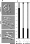
mid2 Δ and spn4 Δ cells have similar cell–cell separation defects. (A) DIC images of representative wild-type (FC937), mid2Δ (FC881), and spn4Δ (FC867) mutant cells grown in rich medium at 30°C to the exponential phase of growth. (B) Numbers of septa in wild-type, mid2Δ, and spn4Δ cells (n > 400). Bar, 10 μm.
Figure 3.
Figure 3.
The contractile ring appears to assemble and close normally in mid2 Δ cells. (a) Wild-type and mid2Δ cells expressing cdc4-GFP were imaged live using confocal time-lapse microscopy. Z-stacks were rendered in three dimensions to produce a cross-sectional view of the ring. Bar, 2 μm. (B) Measurements of five individual ring diameters over time in wild-type and mid2Δ cells.
Figure 4.
Figure 4.
Organization of septin rings is not maintained in mid2 Δ cells. (A) Wild-type (FC937) and mid2Δ (FC881) cells expressing Spn4p-GFP were imaged for GFP fluorescence using confocal three-dimensional time-lapse microscopy. Spn4p-GFP structures were rendered in three dimensions at each time point. Side (0°) and cross-sectional (90°) views of the Spn4p-GFP medial structures are shown in wild-type (left) and mid2Δ (right) at representative time points. Note that in mid2Δ cells, Spn4p-GFP forms a single ring (0 min), an abnormal washer (3–13 min), and then a disc structure (24 min). Bar, 2 μm. (B) mid2Δ cells expressing Spn4p-YFP (green) and the contractile ring marker Cdc12-CFP (red) were imaged on a wide field microscope and rendered in three dimensions. Spn4p-YFP is present on the membrane behind the contractile ring in a washer structure. (C) Summary of the distribution of septins in wild-type and mid2Δ cells during contractile ring closure.
Figure 5.
Figure 5.
Measurement of Spn4p-GFP dynamics using FRAP. (A) Wild-type and mid2Δ cells expressing Spn4p-GFP were photobleached in the zones marked by white rectangles. Recovery of fluorescence intensity was assayed over time (as described in Materials and methods). Representative images are shown. Top panels show cells just before photobleaching. 0 s represents time immediately after photobleaching. (B) Graph of mean t1/2 ± SD of Spn4p-GFP fluorescence recovery in seconds.
Figure 6.
Figure 6.
Mid2p colocalizes with Spn4p. Cells (FC882) expressing Mid2p-YFP (green) and Spn4p-CFP (red) were imaged for YFP and CFP fluorescence. (Top) Cells in late mitosis just after initial assembly of the single septin ring. Some of these cells exhibit septin but not Mid2p staining (left cell). (Middle) Cells during septation with double septin rings. (Bottom) Cells during cell–cell separation and during interphase. Bar, 10 μm.
Figure 7.
Figure 7.
Mid2p localization is dependent on septins and the Mid2p PH domain. (A) spn4Δ cells (FC492) expressing Mid2p-GFP were imaged for GFP fluorescence. Only background fluorescence is observed. (B) Cells expressing only Mid2p-PHΔ were imaged by DIC microscopy. (C) mid2-YFP and mid2-PHΔYFP cells were imaged for YFP fluorescence. Only background fluorescence is observed in the mid2-PHΔYFP cells. Bar, 10 μm.
Figure 8.
Figure 8.
mid1 and mid2 do not show genetic interactions. (A) Wild-type, mid1Δ, mid2Δ, and mid1Δmid2Δ strains were assayed for growth rates and cellular integrity at 30 and 36°C by growth on agar plates containing phloxin, a dye that stains dead cells. Three independent mid1Δmid2Δ strains are represented in the bottom three streaks of each plate. (B) Cells grown in liquid cultures were stained with Calcofluor to visualize septa. (C) Localization of Mid1p-GFP in mid2Δ cells and localization of Mid2p-GFP in mid1Δ cells.

References

    1. Adam, J.C., J.R. Pringle, and M. Peifer. 2000. Evidence for functional differentiation among Drosophila septins in cytokinesis and cellularization. Mol. Biol. Cell. 11:3123–3135. - PMC - PubMed
    1. Altschul, S.F., W. Gish, W. Miller, E.W. Myers, and D.J. Lipman. 1990. Basic local alignment search tool. J. Mol. Biol. 215:403–410. - PubMed
    1. Bahler, J., A.B. Steever, S. Wheatley, Y. Wang, J.R. Pringle, K.L. Gould, and D. McCollum. 1998. a. Role of polo kinase and Mid1p in determining the site of cell division in fission yeast. J. Cell Biol. 143:1603–1616. - PMC - PubMed
    1. Bahler, J., J.Q. Wu, M.S. Longtine, N.G. Shah, A. McKenzie, A.B. Steever, A. Wach, P. Philippsen, and J.R. Pringle. 1998. b. Heterologous modules for efficient and versatile PCR-based gene targeting in Schizosaccharomyces pombe. Yeast. 14:943–951. - PubMed
    1. Balasubramanian, M.K., D. McCollum, and K.L. Gould. 1997. Cytokinesis in fission yeast Schizosaccharomyces pombe. Methods Enzymol. 283:494–506. - PubMed

Publication types

MeSH terms