. 2003 Apr 1;100(7):4322-7.
doi: 10.1073/pnas.0736323100. Epub 2003 Mar 24.

Macula densa cell signaling involves ATP release through a maxi anion channel

Affiliations

Macula densa cell signaling involves ATP release through a maxi anion channel

Phillip Darwin Bell et al. Proc Natl Acad Sci U S A. .

Abstract

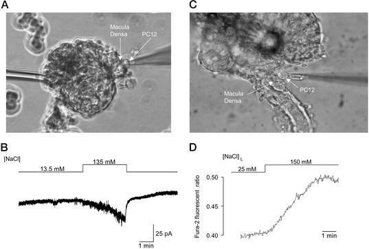
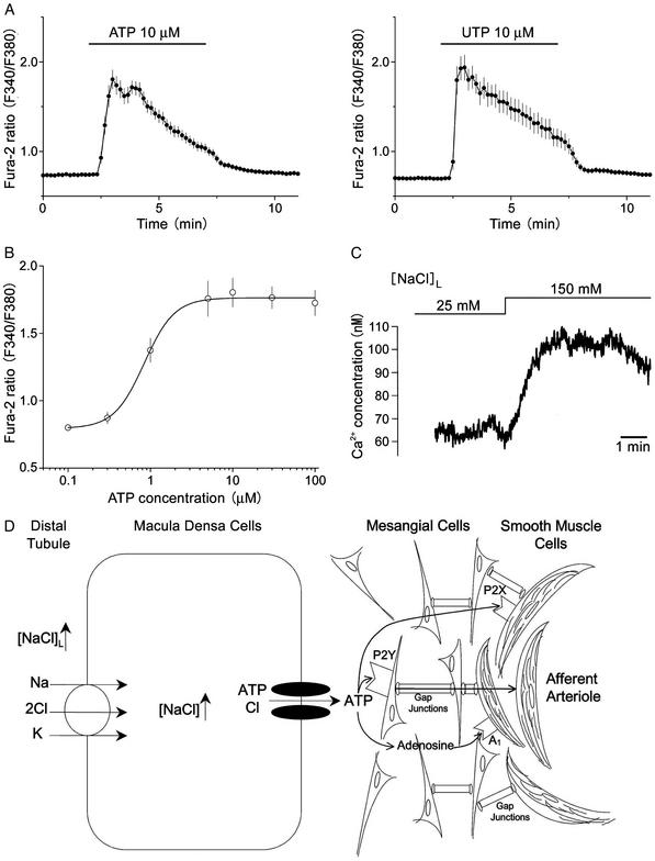
Macula densa cells are unique renal biosensor cells that detect changes in luminal NaCl concentration ([NaCl](L)) and transmit signals to the mesangial cellafferent arteriolar complex. They are the critical link between renal salt and water excretion and glomerular hemodynamics, thus playing a key role in regulation of body fluid volume. Since identification of these cells in the early 1900s, the nature of the signaling process from macula densa cells to the glomerular contractile elements has remained unknown. In patch-clamp studies of macula densa cells, we identified an [NaCl](L)-sensitive ATP-permeable large-conductance (380 pS) anion channel. Also, we directly demonstrated the release of ATP (up to 10 microM) at the basolateral membrane of macula densa cells, in a manner dependent on [NaCl](L), by using an ATP bioassay technique. Furthermore, we found that glomerular mesangial cells respond with elevations in cytosolic Ca(2+) concentration to extracellular application of ATP (EC(50) 0.8 microM). Importantly, we also found increases in cytosolic Ca(2+) concentration with elevations in [NaCl](L), when fura-2-loaded mesangial cells were placed close to the basolateral membrane of macula densa cells. Thus, cell-to-cell communication between macula densa cells and mesangial cells, which express P2Y(2) receptors, involves the release of ATP from macula densa cells via maxi anion channels at the basolateral membrane. This mechanism may represent a new paradigm in cell-to-cell signal transduction mediated by ATP.


Figures

Figure 1
Figure 1
Macula densa-glomerular preparation and NaCl-activated ion channel in the macula densa plasma membrane. (A Left) Schematic diagram of the macula densa–juxtaglomerular apparatus, which illustrates that the macula densa lies within the TAL and is adjacent to the mesangial/arteriolar complex. (Right) Photomicrograph of macula densa–glomerular preparation. The glomerulus was fixed to the bottom of the chamber with a holding pipette (HP). The TAL was removed by dissection, thereby allowing free access of patch pipette (PP) to the macula densa cells. (B) Single-channel recordings from a cell-attached patch on a macula densa cell. An active channel in control Ringer's bath ([NaCl] = 135 mM; Left) was inactivated after removal of NaCl from the bath (Center). Return of NaCl to the bathing solution reactivated the channel (Right). Arrowheads, the closed level of channel current. Arrows, onset of step pulses. The voltage applied to the cell (−Vp) is indicated beside each current trace. Data represent nine similar experiments.
Figure 2
Figure 2
Anion selectivity and ATP permeability of macula densa maxi channel. (A) Single-channel recording from inside-out patches. Shown are current traces in control Ringer's ([NaCl] = 135 mM) solution (Left), low Cl Ringer's solution ([Cl] = 24.5 mM) (Center), and 100 mM ATP solution (Right). The pipette solution always contained 146 mM Cl. (Right) Note the presence of channel activity at negative voltages that represents inward current generated by ATP4− efflux. Arrowheads represent the closed level of channel current, and vertical arrows indicate the step pulse onset. The membrane potential (−Vp) is indicated beside each current trace. (B) Single-channel I-V curves obtained from inside-out patches from macula densa cells. (Left) I-V curves in Ringer's solution containing [NaCl] = 135 mM (n = 12), low Cl (substituted with gluconate; n = 8), or N-methyl-d-glucamine (NMDG; Na+-free; n = 3). (Right) I-V curves in which the bath contained either control Ringer's solution ([NaCl] = 135 mM) or 100 mM ATP solution (n = 7). (C) Expanded trace of single-channel current, presented in A (Right Lower), recorded at −50 mV in 100 mM ATP solution.
Figure 3
Figure 3
NaCl-dependent ATP release from macula densa cells detected by bioassay techniques. (A) The patch pipette with a PC12 cell in whole-cell configuration is positioned on the macula densa plaque. (B) In response to an increase in bath [NaCl], there was activation of a P2X channel. When the PC12 cell was not in contact with the macula densa, no channel activity was present when the bath [NaCl] was increased (data not shown, n = 7). (C) In an isolated perfused TAL in which the glomerulus was partially removed, thereby exposing the basolateral membrane of macula densa plaque, a fura-2-loaded PC12 cell was positioned and held with a holding pipette at the basolateral membrane of the macula densa. (D) In response to an increase in [NaCl]L from 25 to 150 mM, there was an increase in the fura-2 ratio, indicating the activation of P2X receptors and increases in the cytosolic calcium concentration. Data represent 10 similar experiments.
Figure 4
Figure 4
The effects of ATP on cultured mesangial cells. (A) Effects of ATP and UTP on the cytosolic free Ca2+ concentration in single mesangial cells. Each symbol represents the mean values of 47 (for ATP) or 33 (for UTP) experiments, and each bar represents SEM. (B) The concentration-response curve for ATP-induced Ca2+ elevations in mesangial cells. Each symbol represents the mean value of 26–47 experiments, and each bar represents SEM. A curve represents sigmoidal fit with an EC50 value given in the text and with a Hill coefficient of 2.4. The ratio values of basal level and of peak response to 10 μM ATP correspond to the free Ca2+ concentrations of 80.2 ± 4.2 nM (n = 47) and 336.5 ± 25.5 nM (n = 47), respectively. (C) Effect of an increase in [NaCl]L from 25 to 150 mM on the cytosolic free Ca2+ concentration in a single mesangial cell that had been positioned and held with a holding pipette at the basolateral membrane of the macula densa, as shown for PC12 cells in Fig. 3C. The mean Ca2+ concentration increased from 64.7 ± 6.2 to 90.7 ± 9.7 nM (n = 5) in response to an increase in [NaCl]L from 25 to 150 mM. (D) Scheme depicting [NaCl]L-sensitive ATP release from macula densa cells via maxi anion channels at the basolateral membrane, and possible roles of released ATP in mediating signal transduction from macula densa cells to mesangial cells via P2Y receptors and to afferent arteriolar smooth muscle cells via P2X and/or A1 receptors. Signal transduction from mesangial cells to afferent arteriolar smooth muscle cells may take place via gap junctions.

References

    1. Bell P D, Lapointe J-Y. Clin Exp Pharmacol Physiol. 1997;25:541–547. - PubMed
    1. Schnermann J. Am J Physiol. 1998;274:R263–R279. - PubMed
    1. Navar L G, Inscho E W, Majid S A, Imig J D, Harrison-Bernard L M, Mitchell K D. Physiol Rev. 1996;76:425–536. - PubMed
    1. Barajas L. Fed Proc. 1981;40:78–86. - PubMed
    1. Yang T, Park J M, Arend L, Huang Y, Topaloglu R, Pasumarthy A, Praetorius H, Spring K, Briggs J P, Schnermann J. J Biol Chem. 2000;275:37922–37929. - PubMed

Publication types

LinkOut - more resources