. 2003 Apr 1;100(7):3826-31.
doi: 10.1073/pnas.0736456100. Epub 2003 Mar 24.

A metal-binding site in the catalytic subunit of anaerobic ribonucleotide reductase

Affiliations

A metal-binding site in the catalytic subunit of anaerobic ribonucleotide reductase

Derek T Logan et al. Proc Natl Acad Sci U S A. .

Abstract

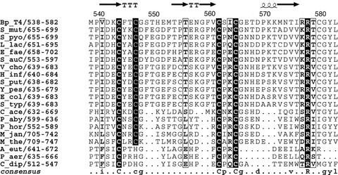
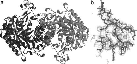
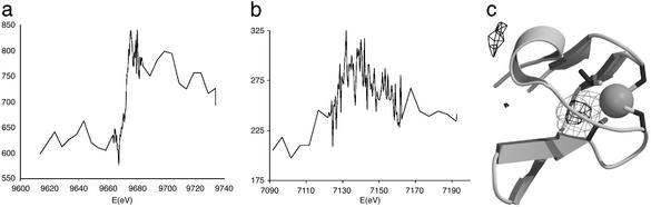
A Zn(Cys)(4) center has been found in the C-terminal region of the crystal structure of the anaerobic class III ribonucleotide reductase (RNR) from bacteriophage T4. The metal center is structurally related to the zinc ribbon motif and to rubredoxin and rubrerythrin. Mutant enzymes of the homologous RNR from Escherichia coli, in which the coordinating cysteines, conserved in almost all known class III RNR sequences, have been mutated into alanines, are shown to be inactive as the result of their inability to generate the catalytically essential glycyl radical. The possible roles of the metal center are discussed in relationship to the currently proposed reaction mechanism for generation of the glycyl radical in class III RNRs.


Figures

Fig 1.
Fig 1.
Sequence alignment of the C-terminal region of the NrdD polypeptide containing the four almost completely conserved cysteine residues. Totally conserved residues are shown in black boxes. The secondary structure as observed in the crystal structure of NrdD from bacteriophage T4 is shown on the top line. T = turn. Figure generated by using ESPRIPT (43). Key to sequence names: Bp_T4, bacteriophage T4; S_mut, Streptococcus mutans; S_pyo, Streptococcus pyogenes; L_lac, Lactococcus lactis; E_fae, Enterococcus faecalis; S_auC, Staphyloccocus aureus COL/NCTC; V_cho, Vibrio cholerae; H_inf, Haemophilus influenzae; S_put, Shewanella putrefaciens; Y_pes, Yersinia pestis; E_col, Escherichia coli; S_typ, Salmonella typhimurium; C_ace, Clostridium acetobutylicum; P_aby, Pyroccocus abyssi; P_hor, Pyrococcus horikoshii; M_jan, Methanococcus jannaschii; M_the, Methanobacterium thermoautotrophicum; A_eut, Alcaligenes eutropha (Ralstonia eutropha); P_aer, Pseudomonas aeruginosa; C_dip, Clostridium difficile.
Fig 2.
Fig 2.
X-band EPR spectra of reduced protein β (90 μM), alone (A) or complexed with wild-type (B) or C665A mutant (C) protein α. Recording conditions: temperature of 10 K, microwave power of 1 mW, and modulation of 1 mT.
Fig 3.
Fig 3.
Structure of the metal-binding domain (MBD) in NrdD and its position in the overall structure. (a) A dimer of NrdD, showing the position of the MBD in relation to the glycyl radical. The monomers are drawn in light and dark gray, respectively. The distance from the metal to the Cα atom of Gly-580 is 28 Å, and the distance between the two metal ions in the dimer is 70 Å. Figs. 3a and 5 were made with MOLSCRIPT (44), Figs. 3b and 4c were made with BOBSCRIPT (45). (b) Electron density for the MBD at 2.45-Å resolution. This is a 2 |Fo| − |Fc| map, with phases from the refined model, contoured at 0.9 σ. An all-atom model for residues 541–574 of NrdD and the metal atom is superimposed.
Fig 4.
Fig 4.
Identity of the metal as shown by absorption edge scans and anomalous difference Fouriers. (a) X-ray fluorescence scan of the K absorption edge of Zn. The fluorescence is given in arbitrary units. In addition, the energy scale is not exactly calibrated due to small errors in the calibration of the synchrotron station. (b) As in a but for the K edge of Fe. (c) Anomalous difference Fourier (thin gray lines) and |Fo| − |Fc| difference Fourier (thick black lines) for Zn. The anomalous map is contoured at 5 σ, the |Fo| − |Fc| map at 4 σ. For the latter map, the Zn atom was omitted from the coordinates, which were then given random displacements of rms 0.1 Å and refined by using REFMAC.
Fig 5.
Fig 5.
Comparison of the fold of the MBD with rubrerythrin and the Zn ribbon motif from transcription factor IID. The coordinating cysteines are labeled. (a) The MBD from NrdD, residues 541–574. (b) Rubrerythrin, C-terminal residues 144–191 (31). (c) Transcription factor IIB N-terminal domain, residues 1–50 from the first model in the PDB file of the NMR structure (26).

References

    1. Jordan A. & Reichard, P. (1998) Annu. Rev. Biochem. 67, 71-98. - PubMed
    1. Sjöberg B.-M. (1997) Struct. Bonding 88, 139-173.
    1. Fontecave M., Mulliez, E. & Logan, D. T. (2002) Prog. Nucleic Acid Res. Mol. Biol. 72, 95-127. - PubMed
    1. Logan D. T., Andersson, J., Sjöberg, B.-M. & Nordlund, P. (1999) Science 283, 1499-1504. - PubMed
    1. Andersson J., Westman, M., Sahlin, M. & Sjöberg, B.-M. (2000) J. Biol. Chem. 275, 19449-19455. - PubMed

Publication types

MeSH terms

LinkOut - more resources