. 2003 Apr 1;100(7):3677-82.
doi: 10.1073/pnas.0737682100. Epub 2003 Mar 24.

Salmochelins, siderophores of Salmonella enterica and uropathogenic Escherichia coli strains, are recognized by the outer membrane receptor IroN

Affiliations

Salmochelins, siderophores of Salmonella enterica and uropathogenic Escherichia coli strains, are recognized by the outer membrane receptor IroN

K Hantke et al. Proc Natl Acad Sci U S A. .

Abstract

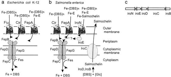
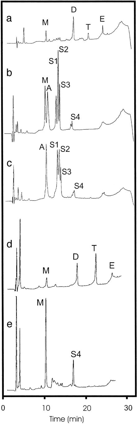
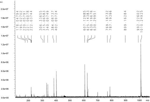
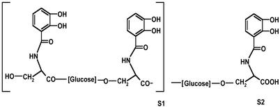
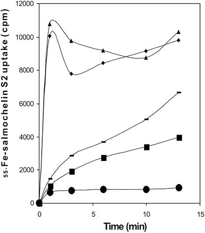
Members of a family of catecholate siderophores, called salmochelins, were isolated by reversed-phase HPLC from Salmonella enterica serotype Typhimurium and structurally characterized by Fourier transform ion cyclotron resonance-MSMS and GC-MS. The tentative structure of salmochelin 1 contained two 2,3- dihydroxybenzoylserine moieties bridged by a glucose residue, bound to the serine hydroxyl group of one moiety and the carboxylate of the second moiety. Salmochelin 2 contained in addition a second glucose residue linked to a third 2,3-dihydroxybenzoylserine moiety. Salmochelins were not produced by an iroBC mutant, which indicated that the IroB protein might be responsible for the glucosyl transfer predicted by sequence similarities to known glycosyltransferases. Uptake experiments with radiolabeled (55)Fe-salmochelin and growth promotion tests with salmochelins showed that the IroN outer membrane receptor, encoded in the iroA locus of S. enterica and uropathogenic Escherichia coli strains, was the main receptor for ferric salmochelin transport.


Figures

Figure 1
Figure 1
Tentative scheme of the catecholate siderophore transport systems of E. coli K-12 (a) and S. enterica (b). In both systems, ferric enterochelin (Fe-E) is transported mainly through FepA across the outer membrane, whereas the linear degradation products, ferric DHBS (DHBS)n (n = 1, 2, 3), are taken up mainly via Fiu and FepA in E. coli. In Salmonella, this has not been studied in detail. However, because of the high similarity of the two systems, the same specificities can be expected. Iron complexes are then transported via the ABC transporter consisting of the binding protein FepB, the membrane components FepD and FepG, and the ATPase FepC through the cytoplasmic membrane. Inside the cell, the Fes protein is required for iron release from the ferric enterochelin complex. In Salmonella, salmochelins are synthesized from glucose and DHBS, possibly with the help of IroD and IroB and secreted by the exporter IroC. IroE might degrade the iron–salmochelin complexes to allow their transport via the ABC transporter FepBDGC. (c) The same gene order is found in the iroA gene locus of S. enterica and uropathogenic E. coli strains (5, 7).
Figure 2
Figure 2
The pooled iron siderophore fraction of E. coli CA46 (ac) eluted from DE52-cellulose with 2 M ammonium chloride was adjusted to pH 2 with H2SO4, extracted with ethyl acetate, and analyzed by HPLC. (a) The ethyl acetate extract contained the linear degradation products monomer DHBS (M), the dimer (DHBS)2 (D), and the trimer (DHBS)3 (T), and traces of the cyclic enterochelin (E) were identified. In the water phase of the ethyl acetate extract (b), the monomer, aerobactin (A), and salmochelins 1–3 (S1–S3) were found. (c) The ammonium chloride eluate of the DE52-cellulose column contained mainly salmochelins 1–3 and aerobactin. (d) The mutant Salmonella H5546 iroBC∷kan produced only enterochelin and the linear degradation products. (e) The mutant Salmonella H5547 iroD∷kan secreted the monomer and S4, which represents an unidentified compound that awaits further characterization.
Figure 3
Figure 3
SORI–CID–MS/MS spectrum of salmochelin 2 and assignment of the resulting fragment ions: m/z 1010.25, [M–H]; m/z 787.19, [M–H–DHB–Ser]; m/z 625.14, [M–H–DHB–Ser–glucose]; m/z 402.10, [M–H–DHB–Ser–glucose–DHB–Ser]; and m/z 240.04, [M–H–DHB–Ser–glucose–DHB–Ser–glucose]. The SORI–CID–MS/MS spectrum of salmochelin 1 (see Fig. 6) has the assigned fragment ions m/z 625.12, [M–H]; m/z 402.09, [M–H–DHB–Ser]; m/z 315.06, [M–H–DHB–Ser–(Ser–H2O)] (rearrangement); and m/z 240.05, [M–H–DBH–Ser–glucose].
Figure 4
Figure 4
Tentative structures of salmochelin 1 (S1) and salmochelin 2 (S2).
Figure 5
Figure 5
Uptake of ferric salmochelin 2 labeled with 55Fe, measured in M9 minimal medium (22) by using the strains S. enterica serotype Stanleyville (⧫), WR1359 iroN (−), WR1361 cir (▴), WR1363 cir iroN (■), and WR1368 cir fepA iroN (●).

References

    1. Earhart C F. In: Escherichia coli and Salmonella. Neidhart F C, editor. Washington, DC: Am. Soc. Microbiol.; 1996. pp. 1075–1090.
    1. Young I G, Gibson F. Methods Enzymol. 1979;56:394–398. - PubMed
    1. Konopka K, Neilands J B. Biochemistry. 1984;23:2122–2127. - PubMed
    1. Bäumler A J, Tsolis R M, van der Velden A W, Stojiljkovic I, Anic S, Heffron F. Gene. 1996;183:207–213. - PubMed
    1. Bäumler A J, Norris T L, Lasco T, Voigt W, Reissbrodt R, Rabsch W, Heffron F. J Bacteriol. 1998;180:1446–1453. - PMC - PubMed

Publication types

MeSH terms

LinkOut - more resources