

doi: 10.1073/pnas.0230529100.
Epub 2003 Mar 24.
Pitx3 is required for development of substantia nigra dopaminergic neurons
Affiliations
- PMID: 12655058
- PMCID: PMC153078
- DOI: 10.1073/pnas.0230529100
Item in Clipboard
Pitx3 is required for development of substantia nigra dopaminergic neurons
Proc Natl Acad Sci U S A.
.
Abstract
Dopaminergic (DA) neurons of substantia nigra in the midbrain control voluntary movement, and their degeneration is the cause of Parkinson's disease. The complete set of genes required to specifically determine the development of midbrain DA subgroups is not known yet. We report here that mice lacking the bicoid-related homeoprotein Pitx3 fail to develop DA neurons of the substantia nigra. Other mesencephalic DA neurons of the ventral tegmental area and retrorubral field are unaltered in their dopamine expression and histological organization. These data suggest that Pitx3-dependent gene expression is specifically required for the differentiation of DA progenitors within the mesencephalic DA system.
Figures

TH immunohistochemistry of adult wt (+/+) (A, C, and E) and ak/ak (−/−) (B, D, and F) ventral midbrain. Coronal sections through rostral (A and B), middle (C and D), and caudal (E and F) levels were stained. TH IR was strong in VTA, SN pars lateralis (SNpl), and A8 of both wt and mutant sections throughout the midbrain but absent in SNpc and SNr. ml, medial lemniscus.
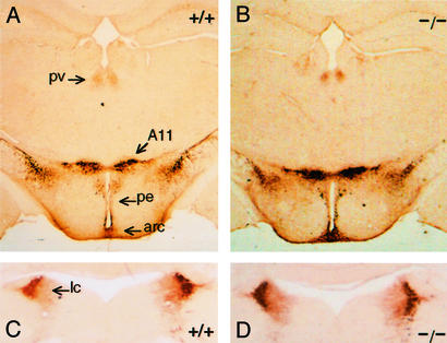
TH immunohistochemistry of adult wt (+/+) and ak/ak (−/−) diencephalon (A and B) and brainstem (C and D). Levels of TH IR in DA neurons of the diencephalon (A and B) and noradrenergic neurons in locus ceruleus (lc) (C and D) were similar in sections from wt and mutant mice. pv, paraventricular thalamic nucleus; pe, periventricular hypothalamic nucleus; arc, arcuate hypothalamic nucleus. A11, caudal thalamus.
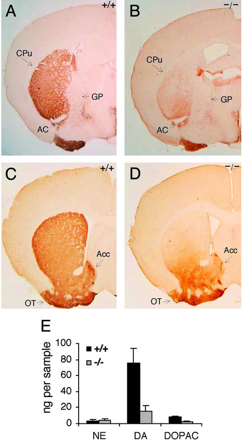
TH immunohistochemistry of adult wt (+/+) (A and C) and ak/ak (−/−) (B and D) striatum. Coronal sections of the entire striatum were analyzed, but sections representative of caudal (A and B) and rostral (C and D) levels are presented. AC, anterior commissure; GP, globus pallidus. Reduced TH staining was most significant in the dorsolateral region of mutant CPu compared with wt mice. TH IR of olfactory tubercle (OT) and accumbens (Acc) was equal among wt and ak sections. (E) NE, DA, and DOPAC levels in micropunches of wt and ak/ak striata were measured by using HPLC. The data are graphed as nanograms of neurotransmitter per micropunch. The tissue volumes in all micropunches were equal. Decreased levels of DA and DOPAC are statistically significant (n = 8, P < 0.001).
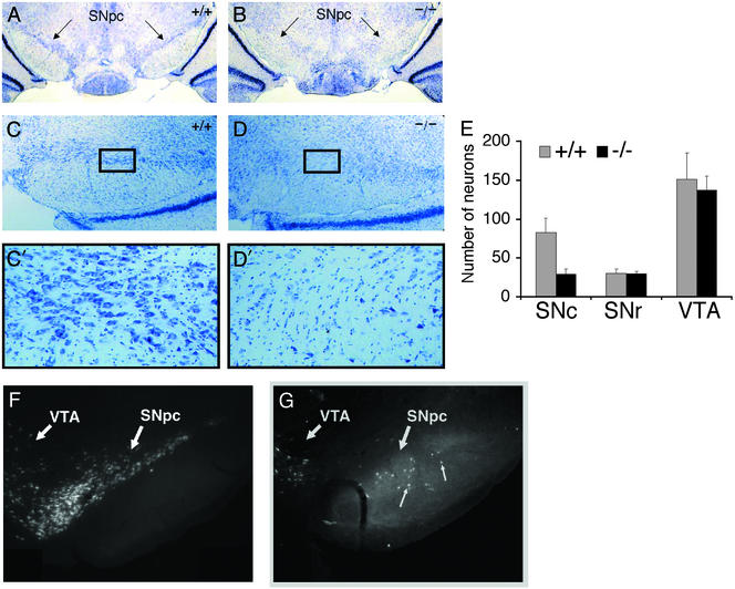
Nissl staining of wt (+/+) (A and C) and ak/ak (−/−) (B and D) adult ventral midbrain. Representative coronal sections are presented to document the decreased cell density in mutant SNpc. Photographs at lower (A and B, ×25; C and D ×100) and higher (C′ and D′, boxed fields, ×400) magnification are provided. (E) Neurons in SNc, SNr, and VTA of wt and mutant Nissl-stained sections were counted. Neurons were distinguished from glial cells based on differences in cell size. Cell counts from four fields (×400 magnification) were averaged and graphed. (F and G) Retrograde labeling of ventral midbrain neurons in wt (F) and ak/ak (G) mice with Fluoro-Gold. Note the absence of cell labeling in mutant SN and aberrant labeling of cells in mutant SNr (small arrows). The neuronal population in the VTA of wt and mutant mice was similar with respect to cell density and location.
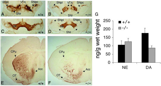
TH immunohistochemistry of newborn (P2) wt (+/+) and ak/ak (−/−) ventral midbrain (A–D) and striata (E and F). Coronal sections through rostral (A and B) and middle (C and D) levels of midbrain were stained. TH IR was strong in the VTA of both wt and mutant mice but absent in mutant SNpc and SNr. ml, medial lemniscus. (E and F) Coronal sections throughout the striata of wt and ak mice were stained, and representative sections are presented. TH IR was reduced significantly in the dorsolateral region of mutant CPu when compared with wt striatum. (G) NE and DA levels in dissected P2 forebrains of wt and ak mice were measured by using HPLC. The data are presented as nanograms of neurotransmitter per gram of dissected tissue. The reduction of DA levels in mutant forebrains is statistically significant (n = 8, P < 0.001).
References
-
- Seeman P, Guan H C, Van Tol H H. Nature. 1993;365:441–445. - PubMed
-
- Schultz W. Neuroscientist. 2001;7:293–302. - PubMed
-
- Damier P, Hirsch E C, Agid Y, Graybiel A M. Brain. 1999;122:1437–1448. - PubMed
-
- Bjorklund A, Lindvall O. In: Classical Transmitters in the CNS: Part I. Bjorklund A, Hokfelt T, editors. Amsterdam: Elsevier Science; 1984. pp. 55–122.
-
- Zetterstrom R H, Solomin L, Jansson L, Hoffer B J, Oson L, Perlmann T. Science. 1997;276:248–250. - PubMed
Publication types
MeSH terms
Substances
Grants and funding
LinkOut - more resources
Full Text Sources
Other Literature Sources
Molecular Biology Databases
