. 2003 Mar 15;23(6):2040-8.
doi: 10.1523/JNEUROSCI.23-06-02040.2003.

Neuronal glutamate uptake Contributes to GABA synthesis and inhibitory synaptic strength

Affiliations

Neuronal glutamate uptake Contributes to GABA synthesis and inhibitory synaptic strength

Gregory C Mathews et al. J Neurosci. .

Abstract

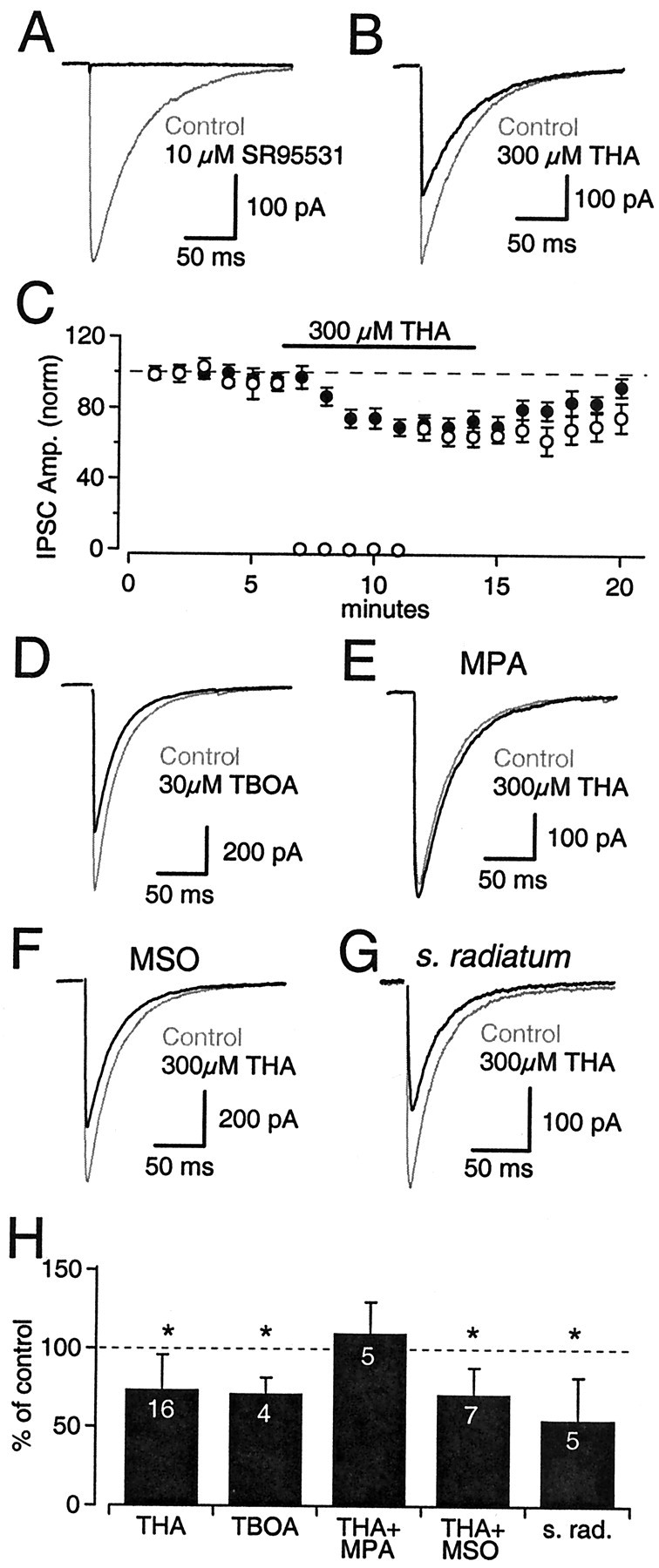
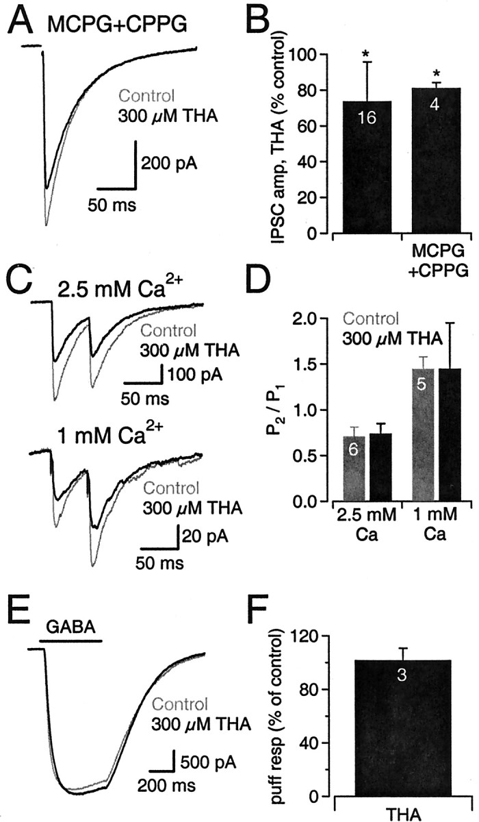
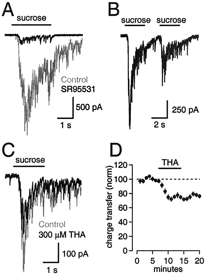
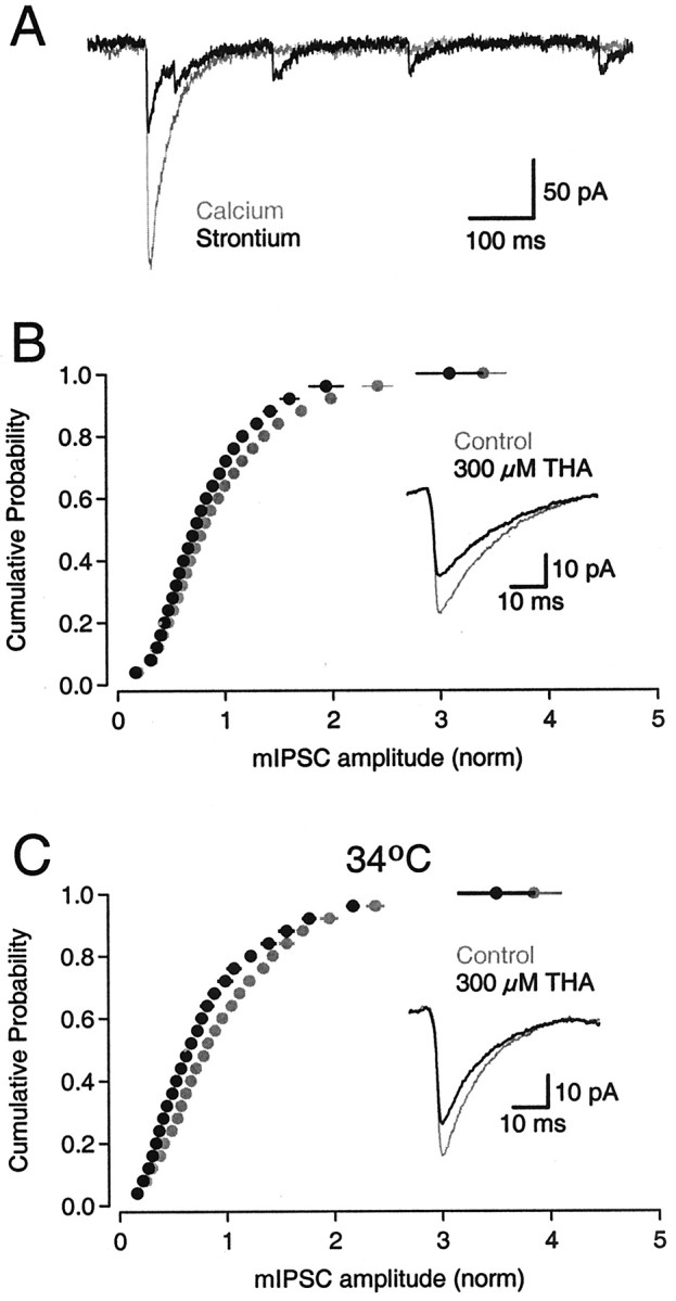
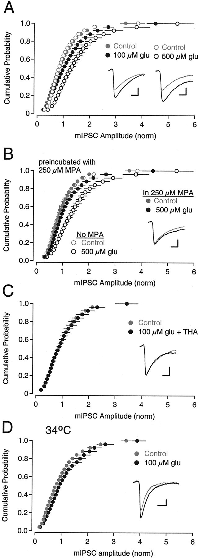
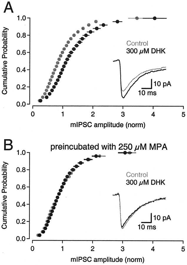
Neurons must maintain a supply of neurotransmitter in their presynaptic terminals to fill synaptic vesicles. GABA is taken up into inhibitory terminals by transporters or is synthesized from glutamate by glutamic acid decarboxylase. Here we report that glutamate transporters supply GABAergic terminals in the hippocampus with glutamate, which is then used to synthesize GABA for filling synaptic vesicles. Glutamate transporter antagonists reduced IPSC and miniature IPSC (mIPSC) amplitudes, consistent with a reduction in the amount of GABA packaged into each synaptic vesicle. This reduction occurred rapidly and independently of synaptic activity, suggesting that modulation of vesicular GABA content does not require vesicle release and refilling. Raising extracellular glutamate levels increased mIPSC amplitudes by enhancing glutamate uptake and, consequently, GABA synthesis. These results indicate that neuronal glutamate transporters strengthen inhibitory synapses in response to extracellular glutamate. This modulation appears to occur under normal conditions and may constitute a negative feedback mechanism to combat hyperexcitability.


Figures

Fig. 1.
Fig. 1.
Inhibition of glutamate transporters reduces electrically evoked IPSCs. A, Evoked IPSC recorded from a CA1 pyramidal cell (Vhold = −60 mV) in control solution (gray) and in the presence of the GABAAR antagonist SR95531 (black).B, Evoked IPSCs in control solution (gray) and in the presence of 300 μm THA (black), a nonselective glutamate transporter antagonist. C, Normalized IPSC amplitudes [Amp. (norm)] during experiments testing the effects of THA (n = 16 cells) with stimulation either continued (filled circles) or suspended (open circles) during wash-in. Error bars indicate SEM.D, IPSC in control solution (gray) and in the presence of 30 μm TBOA (black), a nonselective, nonsubstrate inhibitor of glutamate uptake. E, IPSC in control solution (gray) and in the presence of 300 μm THA (black) after inhibition of GABA synthesis with 250 μm MPA, a GAD inhibitor.F, IPSC in control solution (gray) and in the presence of 300 μm THA (black) after inhibition of astrocytic glutamine synthetase with 1.5 mm MSO.G, IPSCs recorded from a CA1 pyramidal cell after stimulation in stratum radiatum (s. radiatum) in control solution (gray) and in the presence of 300 μm THA (black). H, Summary of effects of glutamate transporter antagonists on IPSC amplitude in various conditions. White numbers in barsindicate the numbers of cells tested. Error bars indicate SD.Asterisks denote a statistically significant difference compared with control (p < 0.05).s. rad., Stratum radiatum.
Fig. 2.
Fig. 2.
THA does not affect release probability or postsynaptic GABAA receptors. A, In the presence of group I and II and group III metabotropic glutamate receptor antagonists (MCPG, 500 μm; and CPPG, 200 μm, respectively), THA still reduced evoked IPSCs.B, Bar graph comparing the effects of THA alone and in the presence of mGluR antagonists. amp, Amplitude.C, Effects of 300 μm THA on IPSCs elicited by a pair of stimuli (50 msec interval). Paired pulse depression was observed in normal (2.5 mm) calcium (top), and paired pulse facilitation was observed in low (1 mm) calcium (bottom). THA (black) reduced IPSC amplitudes compared with control (gray) but did not affect the paired pulse ratio. D, Summary bar graph showing that THA (black bars) affected neither paired pulse depression nor facilitation. E, Currents elicited in a pyramidal cell by puff application of GABA in control solution (gray) and in the presence of 300 μm THA (black; THA was included in the bath and puffer solutions). F, Summary graph showing that THA did not affect GABAA receptors on pyramidal cells. In all bar graphs, white numbers indicate the numbers of cells tested, and error bars indicate SD.
Fig. 3.
Fig. 3.
THA reduces hyperosmotically stimulated release of GABA. A, Postsynaptic current elicited by puff application of control solution with 500 mm sucrose (gray). The puffer was directed toward the pyramidal cell body. The response was almost completely abolished when the GABAA antagonist SR95531 (10 μm) was added to the bath (black). B, The responses to a second puff application of sucrose 1 sec after the first indicated that the releasable pool only partially recovered from depletion by the first puff. C, Response to hyperosmotic stimulation in control solution (gray) and in the presence of 300 μm THA (black).D, Normalized charge transfer during sucrose-evoked IPSCs in the presence of 300 μm THA (n = 9 cells). THA was not included in the sucrose solution. Error bars indicate SEM.
Fig. 4.
Fig. 4.
THA reduces mIPSC amplitudes. A, Electrically evoked IPSCs in normal calcium (gray) and in the same cell after calcium was replaced with strontium (black). In the presence of strontium, a diminished synchronous component of the response was followed by asynchronously released events, which were collected and pooled for each cell under each experimental condition.B, mIPSC amplitudes in control (strontium) solution (gray symbols; n = 1624 events) and in the additional presence of 300 μm THA (black symbols; n = 1491 events). Events in each of 11 cells were normalized (norm) to the mean mIPSC amplitude in control, and cumulative probability histograms were placed into 25 equal bins (p between 0 and 1), allowing events in different cells to be pooled. Error bars indicate cell-to-cell variability (SEM) of the normalized histograms.Inset, Average mIPSC waveforms from a representative cell in control solution (gray) and in the presence of THA (black). C, mIPSC amplitudes in control (strontium) solution (gray symbols; n = 909 events) and in the additional presence of 300 μm THA (black symbols; n = 958 events) recorded at 34°C. Inset, Average mIPSC waveforms from a representative cell in control solution (gray) and in the presence of THA (black).
Fig. 5.
Fig. 5.
Exogenously applied glutamate increases mIPSC amplitudes. A, Addition of 100 μm(black, closed circles; n = 1975 events) or 500 μm (black, open circles;n = 1477 events) glutamate (glu) increased mIPSC amplitude compared with control (gray, closed circles;n = 1990; gray, open circles;n = 1493, respectively). Cumulative probability histograms were constructed as described in the Figure4B legend. Inset, Average mIPSC waveforms from two representative cells in control solution (gray) and in the presence of either 100 μm (black, left) or 500 μm(black, right) glutamate. B, Preincubation with MPA, a GAD inhibitor, reduced the effect of 500 μm glutamate on mIPSC amplitude. The data for 500 μm glutamate from A(open circles) are included for comparison.Inset, Average mIPSC waveforms from a representative cell preincubated in MPA in control solution (gray) and in the presence of 500 μm glutamate (black). C, Three hundred micromolar THA blocked the effect of glutamate on mIPSCs. mIPSCs recorded during coapplication of 300 μm THA and 100 μm glutamate (black symbols;n = 1418 events) were not different compared with control (gray symbols; n = 1088 events). Inset, Average mIPSC waveforms from a representative cell in control solution (gray) and in the presence of 300 μm THA and 100 μm glutamate (black). D, Recorded at 34°C, addition of 100 μm glutamate (black symbols; n = 1177 events) increased mIPSC amplitude compared with control (gray symbols; n = 973 events).Inset, Average mIPSC waveforms from a representative cell in control solution (gray) and in the presence of 100 μm glutamate (black).norm, Normalized.
Fig. 6.
Fig. 6.
Presynaptic glutamate transporters do not exhibit GLT-1 pharmacology. A, Addition of 300 μmDHK (black symbols; n = 1409 events), a selective GLT-1 blocker, increased mIPSC amplitudes compared with control (gray symbols; n= 1455 events). Inset, Average mIPSC waveforms from a representative cell in control solution (gray) and in the presence of DHK (black). B, After preincubation with MPA, 300 μm DHK (black; n = 1096 events) no longer increased mIPSC amplitudes compared with control (gray; n = 790 events).Inset, Average mIPSC waveforms from a representative cell in control solution (gray) and in the presence of DHK (black). norm, Normalized.

Similar articles

Cited by

References

    1. Arnth-Jensen N, Jaboudon D, Scanziani M. Cooperation between independent hippocampal synapses is controlled by glutamate uptake. Nat Neurosci. 2002;5:325–331. - PubMed
    1. Arriza JL, Fairman WA, Wadiche JI, Murdoch GH, Kavanaugh MP, Amara SG. Functional comparisons of three glutamate transporter subtypes cloned from human motor cortex. J Neurosci. 1994;14:5559–5569. - PMC - PubMed
    1. Asztely F, Erdemli G, Kullmann DM. Extrasynaptic glutamate spillover in the hippocampus: dependence on temperature and the role of active glutamate uptake. Neuron. 1997;18:281–293. - PubMed
    1. Bergles DE, Jahr CE. Synaptic activation of glutamate transporters in hippocampal astrocytes. Neuron. 1997;19:1297–1308. - PubMed
    1. Brasnjo G, Otis TS. Neuronal glutamate transporters control activation of postsynaptic metabotropic glutamate receptors and influence cerebellar long-term depression. Neuron. 2001;31:607–616. - PubMed

Publication types

MeSH terms

LinkOut - more resources