Comparative Study
. 2003 Mar 15;23(6):2086-92.
doi: 10.1523/JNEUROSCI.23-06-02086.2003.

The binding of 2-(4'-methylaminophenyl)benzothiazole to postmortem brain homogenates is dominated by the amyloid component

Affiliations
Comparative Study

The binding of 2-(4'-methylaminophenyl)benzothiazole to postmortem brain homogenates is dominated by the amyloid component

William E Klunk et al. J Neurosci. .

Abstract

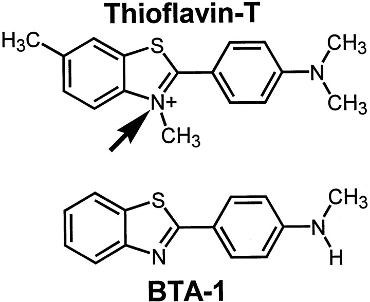
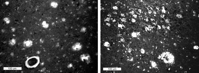
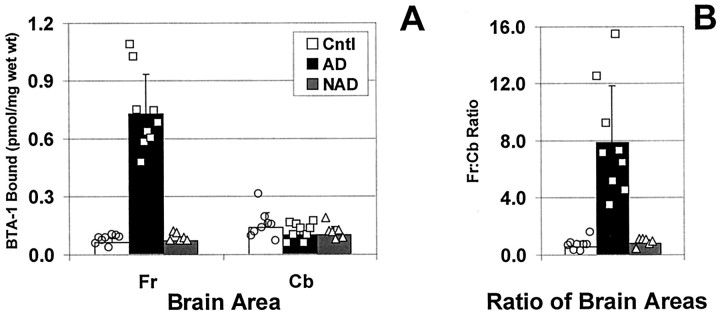
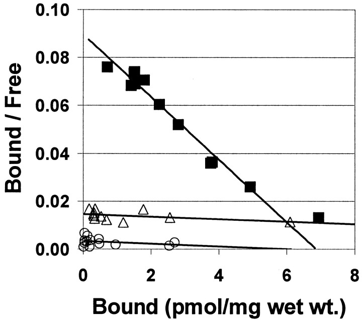
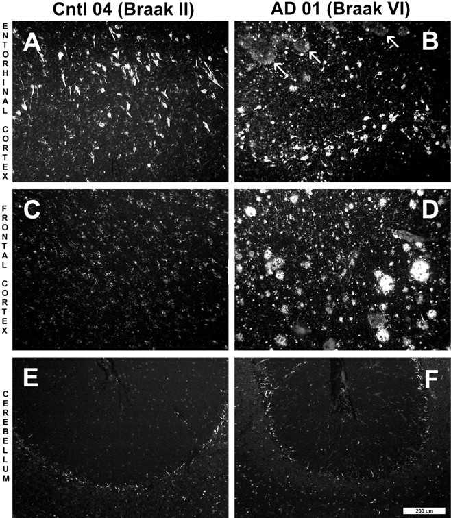
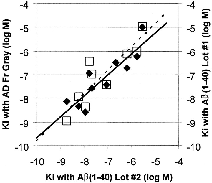
2-(4'-methylaminophenyl)benzothiazole (BTA-1) is an uncharged derivative of thioflavin-T that has high affinity for Abeta fibrils and shows very good brain entry and clearance. In this study, we asked whether BTA-1, at concentrations typical of those achieved during positron emission tomography (PET) studies, could specifically bind to amyloid deposits in the complex milieu of human brain or whether amyloid binding was overshadowed by nonspecific binding, found even in brains that did not contain amyloid deposits. We quantitatively assessed [3H]BTA-1 binding to crude homogenates of postmortem brain obtained from nine Alzheimer's disease (AD) subjects, eight controls, and six subjects with non-AD dementia. BTA-1 binding was >10-fold higher in AD brain, and the majority (94%) of the binding was specific (displaceable). High-affinity [3H]BTA-1 was observed only in AD brain gray matter and was not present in control brain gray matter, AD brain white matter, or cerebellum. The K(d) of [3H]BTA-1 for binding to AD brain (5.8 +/- 0.90 nm) was very similar to the K(d) for binding to synthetic Abeta fibrils. In addition, the K(i) of various BTA analogs for inhibition of [3H]BTA-1 binding to AD brain homogenates was very similar to their K(i) for inhibition of [3H]BTA-1 binding to synthetic Abeta fibrils. Nanomolar concentrations of [3H]BTA-1 did not appear to bind to neurofibrillary tangles. Finally, BTA-1 did not appear to bind significantly to common neuroreceptors or transporter sites. These data suggest that the binding of BTA-1 to AD brain is dominated by a specific interaction with Abeta amyloid deposits.


Figures

Fig. 1.
Fig. 1.
Chemical structures of thioflavin-T and BTA-1. The uncharged compound, BTA-1, differs from thioflavin-T by the lack of three methyl groups, including the methyl group imparting the positive charge to the quaternary heterocyclic nitrogen of thioflavin-T (arrow).
Fig. 2.
Fig. 2.
Paraffin sections (8 μm) from AD brain stained with 100 nm BTA-1. Left panel shows plaques and cerebrovascular amyloid in the frontal cortex. Right panel shows plaques and neurofibrillary tangles in the entorhinal cortex. Scale bar, 100 μm. Most smaller bright spots are residual lipofuscin autofluorescence.
Fig. 3.
Fig. 3.
A, Comparison of [3H]BTA-1 binding to homogenates from control (open bars, circles), AD (filled bars, squares), and NAD brain (hatched bars, triangles) frontal gray (Fr) or cerebellum (Cb).B, Ratio of [3H]BTA-1 binding to the Fr and Cb for each individual brain. Bars represent the mean, and error bars represent the SD. Also shown are the individual data points.
Fig. 4.
Fig. 4.
Scatchard plots showing the binding of [3H]BTA-1 to homogenates from AD frontal gray matter (▪) and underlying frontal white matter from the same AD brain (▵). In this AD frontal gray matter homogenate, theKd was 4.4 nm and theBmax was 6.9 pmol/mg wet weight. In the frontal white matter immediately underlying this gray matter sample, the Kd was 111 nm and theBmax was 27 pmol/mg wet weight. Also shown is a Scatchard plot showing the binding of [3H]BTA-1 to a homogenate from control brain frontal gray (○) in which the Kd was 180 nm and the Bmax was 5.9 pmol/mg wet weight.
Fig. 5.
Fig. 5.
Paraffin sections (8 μm) from the entorhinal cortex (A, B), frontal cortex (C, D), and cerebellum (E,F) of a Braak stage II control brain (A, C, E, Cntl 04) and a Braak stage VI AD brain (B,D, F, AD 01) stained with X-34. X-34 is a highly sensitive, fluorescent Congo red derivative that intensely stains plaques, NFTs, and amyloid deposits in general (Styren et al., 2000). Marked atrophy of AD tissue was notable in all regions. In the entorhinal cortex of the control subject (A), frequent numbers of NFTs were seen, whereas there were no amyloid deposits. In the AD case (B), there also were frequent NFTs along with diffuse amyloid deposits (arrows). X-34-positive NFTs and amyloid plaques were absent from the frontal cortex of the control case (C), whereas they were abundant in AD (D). There was no detectable X-34 staining of plaques or NFT in control (E) and AD (F) cerebellum. Control frontal cortex (C) and cerebellum from both control and AD (E, F) show small stellate cells that stain with X-34. Scale bar, 200 μm.
Fig. 6.
Fig. 6.
Comparison of the Kivalues of 10 BTA-1 derivatives for inhibition of [3H]BTA-1 binding to Aβ(1–40) fibrils and homogenates of AD frontal gray (filled diamonds,solid line). Also shown is a comparison of the same data for Aβ(1–40) fibrils compared with Kivalues previously determined in a separate, older lot of Aβ(1–40) fibrils (open squares, dashed line).

Similar articles

Cited by

References

    1. Bard F, Cannon C, Barbour R, Burke RL, Games D, Grajeda H, Guido T, Hu K, Huang J, Johnson-Wood K, Kholodenko D, Lee M, Lieberburg I, Motter R, Nguyen M, Soriano F, Vasquez N, Weiss K, Welch B, Schenk D, Yednock T. Peripherally administered antibodies against amyloid β-peptide enter the central nervous system and reduce pathology in a mouse model of Alzheimer disease. Nat Med. 2000;6:916–919. - PubMed
    1. Bennett JP, Yamamura HI. Neurotransmitter, hormone, or drug receptor binding methods. In: Yamamura HI, Enna SJ, Kuhar MJ, editors. Neurotransmitter receptor binding. Raven; New York: 1985. pp. 61–89.
    1. DeMattos RB, Bales KR, Cummins DJ, Dodart JC, Paul SM, Holtzman DM. Peripheral anti-A beta antibody alters CNS and plasma A beta clearance and decreases brain A beta burden in a mouse model of Alzheimer's disease. Proc Natl Acad Sci USA. 2001;98:8850–8855. - PMC - PubMed
    1. Goate A, Chartier-Harlin MC, Mullan M, Brown J, Crawford F, Fidani L, Giuffra L, Haynes A, Irving N, James L, Mant R, Newton P, Rooke K, Roques P, Talbot C, Pericak-Vance M, Roses A, Williamson R, Rossor M, Owen M, Hardy J. Segregation of a missense mutation in the amyloid precursor protein gene with familial Alzheimer's disease. Nature. 1991;349:704–706. - PubMed
    1. Goedert M, Spillantini MG. Tau mutations in frontotemporal dementia FTDP-17 and their relevance for Alzheimer's disease. Biochim Biophys Acta. 2000;1502:110–121. - PubMed

Publication types

MeSH terms