. 2003 Mar 15;23(6):2112-21.
doi: 10.1523/JNEUROSCI.23-06-02112.2003.

Ischemic insults derepress the gene silencer REST in neurons destined to die

Affiliations

Ischemic insults derepress the gene silencer REST in neurons destined to die

Agata Calderone et al. J Neurosci. .

Abstract

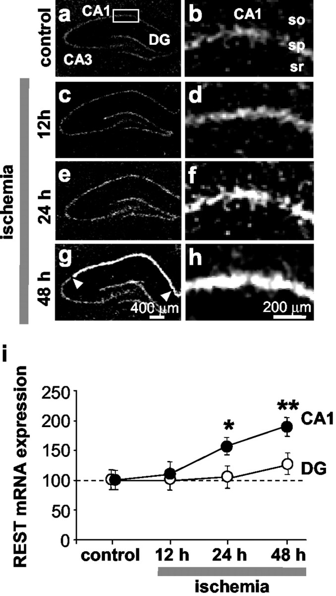
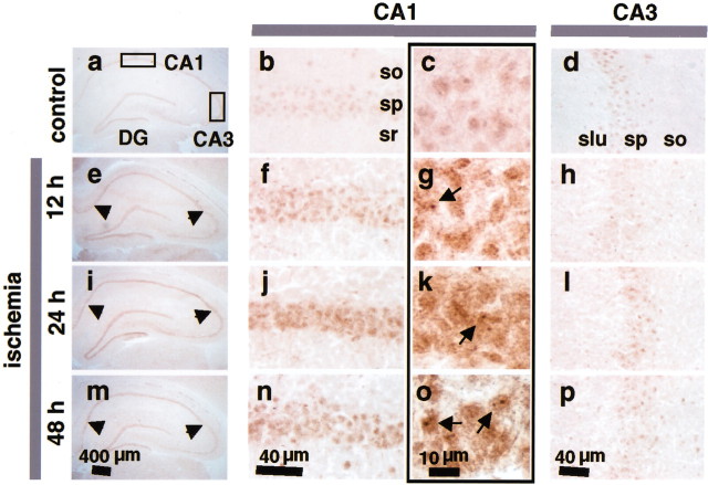
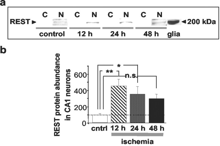
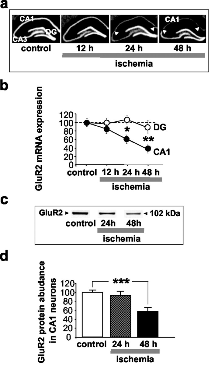
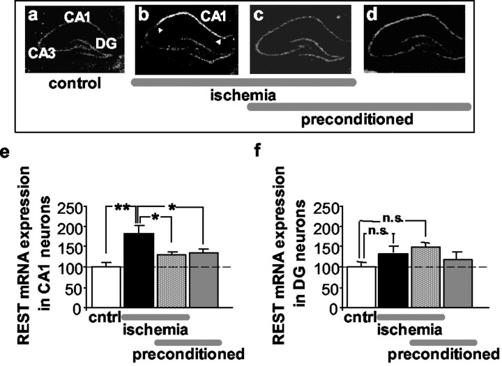
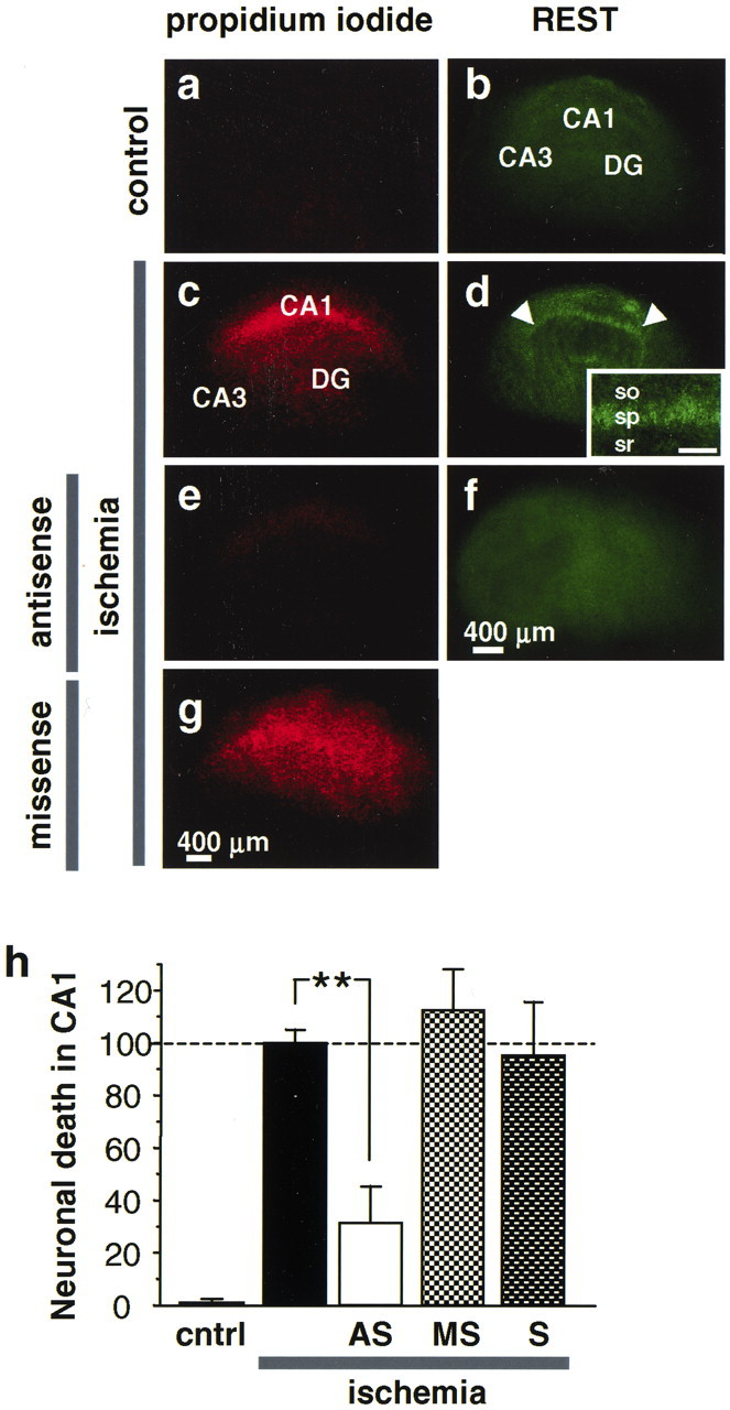
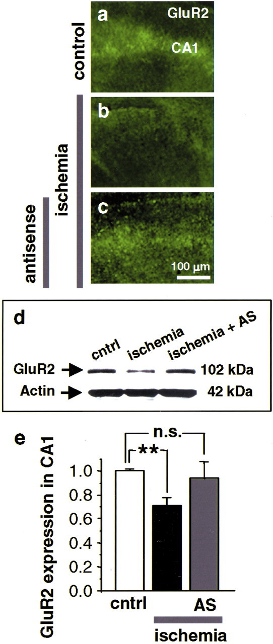
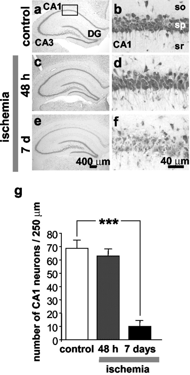
A subset of genes implicated in genetic and acquired neurological disorders encode proteins essential to neural patterning and neurogenesis. The gene silencing transcription factor neuronal repressor element-1 silencing transcription factor (REST)/neuron-restrictive silencer factor (NRSF) plays a critical role in elaboration of the neuronal phenotype. In neural progenitor and non-neural cells, REST acts by repression of a subset of neural genes important to synaptic plasticity and synaptic remodeling, including the AMPA receptor (AMPAR) subunit GluR2. Here we show that global ischemia triggers REST mRNA and protein expression. REST suppresses GluR2 promoter activity and gene expression in neurons destined to die. Because the GluR2 subunit governs AMPAR Ca2+ permeability, these changes are expected to have profound effects on neuronal survival. In keeping with this concept, acute knockdown of the REST gene by antisense administration prevents GluR2 suppression and rescues post-ischemic neurons from ischemia-induced cell death in an in vitro model. To our knowledge, our study represents the first example of ischemia-induced induction of a master transcriptional regulator gene and its protein expression critical to neural differentiation and patterning in adult neurons. Derepression of REST is likely to be an important mechanism of insult-induced neuronal death.


Figures

Fig. 1.
Fig. 1.
Global ischemia increases REST mRNA expression in CA1 pyramidal neurons. ah, Film autoradiograms of REST mRNA expression detected by in situ hybridization with “riboprobe 11” (see Material and Methods) of brain sections at the level of the dorsal hippocampus from control (a, b; n = 3) and experimental rats at 12 hr (c, d;n = 9), 24 hr (e,f; n = 8), and 48 hr (g, h; n = 12) after global ischemia. i, Quantitative analysis of REST mRNA expression in the CA1 (CA1) pyramidal cell layer (●) and the dentate gyrus (DG) granule cell layer (○). At 24 hr after ischemia, REST mRNA expression was increased significantly in the CA1 pyramidal cell layer and slightly, but not significantly, increased in the DG granule cell layer. At 48 hr, REST was dramatically increased in the CA1 (arrowheads), with significantly lower expression in DG and CA3 (data not analyzed, but see autoradiograms). Experiments performed with “riboprobe 4” (see Material and Methods) showed similar results. Mean optical density values are reported after normalization to the control value for the corresponding subfield. Statistical significance was assessed by ANOVA followed by Newman–Keuls test (*p < 0.05; **p < 0.01). Scale bars: a,c, e, g, 400 μm; b,d, f, h, 200 μm.so, Stratum oriens; sp, stratum pyramidale;sr, stratum radiatum.
Fig. 2.
Fig. 2.
Ischemia increases REST protein expression in nuclei of CA1 pyramidal neurons. Coronal sections of rat brain at the level of the dorsal hippocampus were probed with the p73 antibody, a polyclonal antibody directed to the N-terminal DNA binding domain of REST. Representative sections are shown from control (ad) and experimental animals at 12 hr (eh), 24 hr (il), and 48 hr (mp) after global ischemia (n = 5 per treatment group). REST immunolabeling in the entire hippocampus is shown at low magnification (first column), in the CA1 at intermediate (second column) and high magnification (third column), and in the CA3 at intermediate magnification (fourth column). Arrowheads point to the CA1 pyramidal cell layer (e, i,m). Arrows point to the nuclei of post-ischemic CA1 neurons (g, k,o). so, Stratum oriens;sp, stratum pyramidale; sr, stratum radiatum; slu, stratum lucidum. Scale bars:a, e, i, m, 400 μm; b, d, f,h, j, l, n,p, 40 μm; c, g,k, o, 10 μm.
Fig. 3.
Fig. 3.
Ischemia increases REST protein abundance in CA1 cell nuclei. a, b, REST expression in cytosolic (C) and nuclear (N) protein samples isolated from CA1 of control and ischemic animals, as assessed by Western blot analysis.a, Representative Western blots probed with the N-terminal antibody (see Material and Methods); b, REST abundance for protein samples isolated from the CA1 of control and experimental rats at 12, 24, and 48 hr after global ischemia (n = 5 per time point). Equal loading was performed to ensure that the same amount of protein was loaded in each lane. REST protein was at low abundance in samples of control CA1. Ischemia increased REST abundance in CA1 nuclei at 12, 24, and 48 hr after insult. A sample from cultured rat brain astrocytes was used as a positive control. Bars represent means ± SEMs. Statistical significance was assessed by means of an ANOVA followed by Newman–Keuls test (*p < 0.05; **p < 0.01).
Fig. 4.
Fig. 4.
Global ischemia reduces acetylated histone proteins H3 and H4 over the GluR2 promoter. Microdissected tissues from the CA1 (a) and CA3 (b) of control and experimental animals at 24 and 48 hr after ischemia were subjected to chromatin immunoprecipitation with antibodies against acetylated H3 and H4 as described in Material and Methods. DNA was purified from immunoprecipitates and subjected to amplification by PCR with primers directed to the core region of the GluR2 promoter.a, CA1, first lane, 100 and 200 bp size markers labeled in c; second lane, as expected, the size of the PCR product from genomic DNA (35 cycles) is 147 bp and is bracketed on either side by size markers. Omission of the histone antibodies showed negligible PCR product (third and fourth lanes). Signal was prominent in control CA1 and greatly reduced at 24 and 48 hr after ischemia. Eachlane represents a different animal. b, CA3. Signal was prominent in both control and post-ischemic CA3.c, A titration of genomic DNA shows that intensities of PCR bands are proportional to the amount of input DNA (M, size markers). Ischemia dramatically reduced the abundance of GluR2 promoter associated with acetylated histones H3 and H4 in samples from CA1, but not CA3, at 48 after global ischemia. no Ab, Primary antibody omitted.
Fig. 5.
Fig. 5.
Global ischemia suppresses GluR2 mRNA and protein expression in CA1. a, Film autoradiograms of GluR2 mRNA expression, as detected by in situ hybridization, in the hippocampus of control (n = 4) and experimental rats at 12 hr (n = 8), 24 hr (n = 8), and 48 hr (n = 12) after global ischemia.b, Quantitative analysis of GluR2 mRNA expression in the pyramidal cell layer of the CA1 (CA1, ●) and in the granule cell layer of the dentate gyrus (DG, ○). Global ischemia induced a marked suppression of GluR2 mRNA expression specifically in the pyramidal neurons of the CA1 at 24 hr; suppression was maximal at 48 hr. No changes were detected in the DG or CA3. Mean optical densities are reported after normalization to the corresponding control value for a given region as indicated in Materials and Methods. Statistical significance was assessed by ANOVA followed by Newman–Keuls test (*p < 0.05; **p < 0.01). c, Representative Western blots probed with a monoclonal antibody against a sequence within the N-terminal domain of the GluR2 subunit; d, relative GluR2 subunit abundance (defined as the ratio of band densities of experimental vs control samples) for protein samples isolated from the CA1 of control (n = 4) and experimental rats at 24 hr (n = 6) and 48 hr (n = 5) after ischemia. GluR2 abundance was determined from band densities for GluR2 after normalization to the band densities for actin, which served as a loading control. Relative GluR2 subunit abundance was markedly decreased in CA1 at 48 hr. Bars represent means ± SEMs. Statistical significance was assessed by means of the Student's unpaired t test (***p < 0.001).
Fig. 6.
Fig. 6.
Ischemic preconditioning markedly attenuates ischemia-induced expression of REST in CA1.ad, Film autoradiograms of REST mRNA expression detected by in situ hybridization with riboprobe 11 in the hippocampus of rats subjected to sham operation (a), global ischemia (b), ischemic preconditioning followed by global ischemia (c), and preconditioning followed by sham operation (d) at 48 hr after the last reperfusion. Quantitative analyses of REST mRNA expression in the pyramidal cell layer of the CA1 (e) and the granule cell layer of the DG (f) for the four treatment groups. Global ischemia increased rREST mRNA expression in the hippocampal CA1 at 48 hr (n = 12) relative to that of controls (n = 3). Ischemic preconditioning, followed by global ischemia (n = 6), and ischemic preconditioning alone (n = 6) induced modest, but not statistically significant, increases in REST expression in the CA1. No significant changes were observed in the resistant DG. Mean optical densities are reported after normalization to the corresponding control value for a given region as indicated in Material and Methods. Bars represent means ± SEMs. Statistical significance was assessed by ANOVA followed by Newman–Keuls test (*p < 0.05; **p < 0.01). DG, Dentate gyrus;cntrl, control; n.s., not significant.
Fig. 7.
Fig. 7.
REST antisense blocks REST expression and rescues CA1 neurons from OGD-induced death. ag, Propidium iodide labeling of organotypically cultured hippocampal slices. a, Control slice. c, Ischemic slice at 72 hr after OGD (45 min). The pyramidal cell layer of CA1 exhibits intense propidium iodide labeling, indicative of cell death.e, REST antisense (20 μm, administered immediately after OGD) prevents neuronal death. g, REST missense (20 μm, administered immediately after OGD) was without effect. OGD caused extensive neuronal death in the pyramidal cell layer in CA1; REST antisense protected ∼70% of CA1 neurons.b, d, OGD induced REST expression in CA1 pyramidal neurons, as assessed by immunofluorescence detected with the N-terminal antibody at 16 hr. f, REST antisense suppressed ischemia-induced REST expression. h, Quantitation of cell death. Propidium iodide fluorescence in the CA1 pyramidal cell layer, expressed as percentage of that in control slices at 72 hr after OGD. **p < 0.01. Scale bars:ag, 400 μm; inset, 100 μm. cntrl, Control; AS, antisense;MS, missense; S, sense.
Fig. 8.
Fig. 8.
REST antisense blocks GluR2 suppression.ac, GluR2 immunofluorescence in the CA1 of organotypically cultured hippocampal slices at 42 hr after OGD (n = 4). a, Control slice. GluR2 immunofluorescence is intense in the CA1 pyramidal cell layer.b, OGD induces GluR2 suppression in CA1 pyramidal neurons. c, REST antisense (20 μm, administered immediately after OGD) prevents ischemia-induced GluR2 suppression. d, Representative Western blot of samples from the CA1 of cultured slices probed with a GluR2 antibody. OGD decreased GluR2 protein expression in CA1 at 48 hr (OGD lane) (n = 9). REST antisense (20 μm, administered immediately after OGD) blocked GluR2 downregulation (OGD + AS lane) (n = 3). REST sense (20 μm, administered immediately after OGD) was without effect (OGD + S). e, Quantitation of GluR2 protein expression, assessed by Western blot analysis. Band densities were normalized to those for actin. **p < 0.01. Scale bar: (in c) ac, 100 μm.
Fig. 9.
Fig. 9.
Global ischemia induces selective, delayed neuronal death in hippocampal CA1. af, Toluidine blue staining of coronal brain sections at the level of the dorsal hippocampus from control (a, b) (n = 5) and experimental male rats subjected to global ischemia at 48 hr (c, d) (n = 9) and 7 d (e,f) (n = 5) after ischemia. Control animals were killed at 7 d after sham operation. At 48 hr after global ischemia, there was no histologically detectable neuronal death in any hippocampal subfield. At 7 d after ischemia, the pyramidal cell layer of CA1 exhibited dramatic loss of neurons, whereas CA3 and dentate gyrus showed no damage. Scale bars: a,c, e, 400 μm; b,d, f, 40 μm. g, Quantitation of cell counts from brain sections illustrated inaf. To assess hippocampal injury, the number of surviving neurons per 250 μm length in the pyramidal cell layer of the medial CA1 was counted under a light microscope at 40× magnification in sections. Neuronal counts from a minimum of four microscopic sections per animal were analyzed; comparisons among group means were made using the Student's t test (***p < 0.001).

References

    1. Andria ML, Simon EJ. Identification of a neurorestrictive suppressor element (NRSE) in the human mu-opioid receptor gene. Brain Res Mol Brain Res. 2001;91:73–80. - PubMed
    1. Bahn S, Mimmack M, Ryan M, Caldwell MA, Jauniaux E, Starkey M, Svendsen CN, Emson P. Neuronal target genes of the neuron-restrictive silencer factor in neurospheres derived from fetuses with Down's syndrome: a gene expression study. Lancet. 2002;359:310–315. - PubMed
    1. Ballas N, Battaglioli E, Atouf F, Andres ME, Chenoweth J, Anderson ME, Burger C, Moniwa M, Davie JR, Bowers WJ, Federoff HJ, Rose DW, Rosenfeld MG, Brehm P, Mandel G. Regulation of neuronal traits by a novel transcriptional complex. Neuron. 2001;31:353–365. - PubMed
    1. Bessis A, Salmon AM, Zoli M, Le Novere N, Picciotto M, Changeux JP. Promoter elements conferring neuron-specific expression of the beta 2-subunit of the neuronal nicotinic acetylcholine receptor studied in vitro and in transgenic mice. Neuroscience. 1995;69:807–819. - PubMed
    1. Bowie D, Mayer ML. Inward rectification of both AMPA and kainate subtype glutamate receptors generated by polyamine-mediated ion channel block. Neuron. 1995;15:453–462. - PubMed

Publication types

MeSH terms