. 2003 Mar 15;23(6):2161-9.
doi: 10.1523/JNEUROSCI.23-06-02161.2003.

Phospholemman, a single-span membrane protein, is an accessory protein of Na,K-ATPase in cerebellum and choroid plexus

Affiliations

Phospholemman, a single-span membrane protein, is an accessory protein of Na,K-ATPase in cerebellum and choroid plexus

Marina S Feschenko et al. J Neurosci. .

Abstract

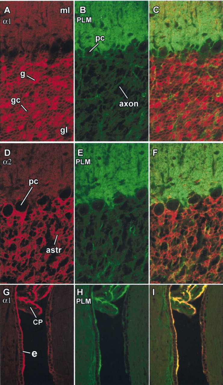
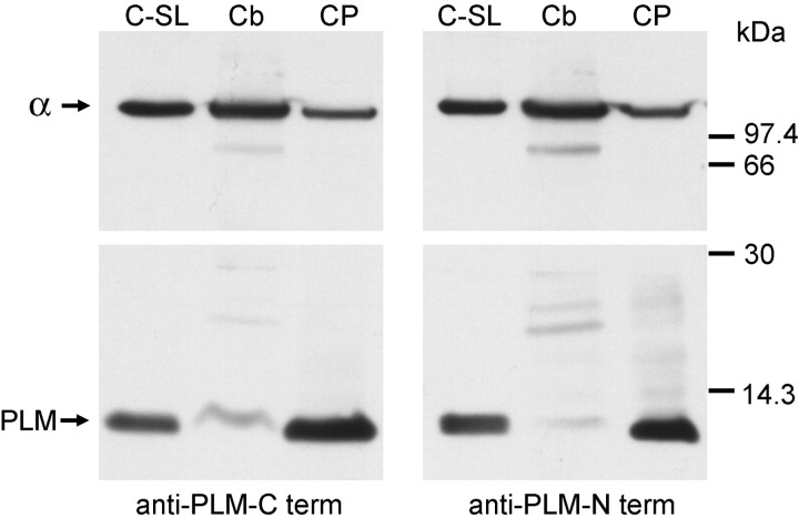
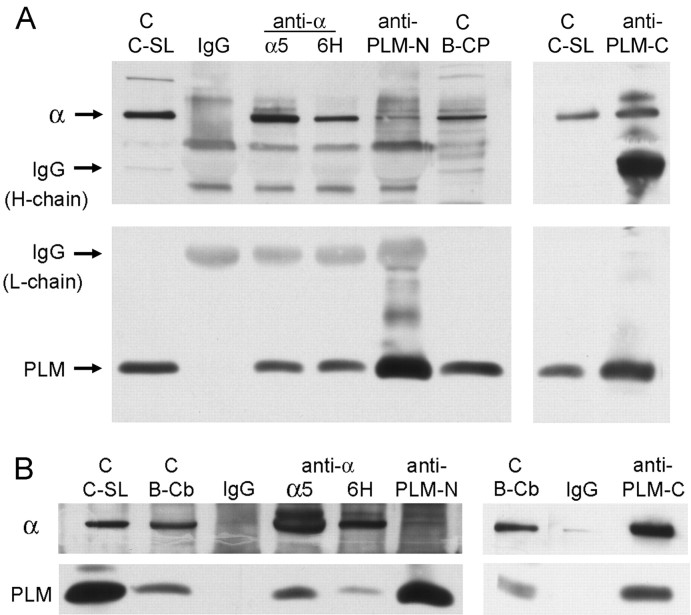
Phospholemman (FXYD1) is a homolog of the Na,K-ATPase gamma subunit (FXYD2), a small accessory protein that modulates ATPase activity. Here we show that phospholemman is highly expressed in selected structures in the CNS. It is most abundant in cerebellum, where it was detected in the molecular layer, in Purkinje neurons, and in axons traversing the granule cell layer. Phospholemman was particularly enriched in choroid plexus, the organ that secretes CSF in the ventricles, where it colocalized with Na,K-ATPase in the apical membrane. It was also enriched, with Na,K-ATPase, in certain tanycytes or ependymal cells of the ventricle wall. Two different experimental approaches demonstrated that phospholemman physically associated with the Na,K-ATPase in cerebellum and choroid plexus: the proteins copurified after detergent treatment and co-immunoprecipitated from solubilized crude membranes using either anti-phospholemman or anti-Na,K-ATPase antibodies. Phospholemman antibodies precipitated all three Na,K-ATPase alpha subunit isoforms (alpha1-alpha3) from cerebellum, indicating that the interaction is not specific to a particular alpha isoform and consistent with the presence of phospholemman in both neurons and glia. Antibodies against the C-terminal domain of phospholemman reduced Na,K-ATPase activity in vitro without effect on Na+ affinity. At least two other FXYD family members have been detected in the CNS, suggesting that additional complexity of sodium pump regulation will be found.


Figures

Fig. 1.
Fig. 1.
Alignment of phospholemman and the γ subunit. The sequences of human phospholemman and γa are compared to display their similarity. One gap was allowed in phospholemman to allow alignment of three identical amino acids in the N-terminal region. The leader sequence of phospholemman and the alternatively spliced N terminus of γ are underlined anditalicized. Following these two structures, the homology begins. As shown previously (Sweadner and Rael, 2000), there is a set of highly conserved amino acids that characterizes the entire FXYD family; these are marked with circles.Lines mark additional residues that are identical between phospholemman and γ. The known phosphorylation sites in the phospholemman C terminus are indicated; this segment has no counterpart in γ.
Fig. 2.
Fig. 2.
Phospholemman immunoreactivity in adult rat brain. A section of rat brain (without the olfactory bulb) stained with antibody against the C-terminal end of phospholemman, PLM-C1, is shown. The brightest immunoreactivity was in the cerebellar molecular layer (ml) and in the choroid plexus (CP), whereas the cerebellar granular layer (gl) was almost unstained. Stained choroid plexus in (left to right) lateral, third, and fourth ventricles can be seen, as well as in presumptive tanycytes of the ependyma of the lateral ventricle (asterisk). Occasional folds in the section produced irregular brighter lines. The image is a montage of many individual confocal images.
Fig. 3.
Fig. 3.
Phospholemman immunoreactivity in cerebellar cortex, choroid plexus, and ventricular wall. A–C, Cerebellar cortex double-labeled for phospholemman (green), the α1 subunit of Na,K-ATPase (red), and the combined images. ml, Molecular layer; gl, granular layer. Immunoreactivity for α1 in the glomeruli (g) in the granular layer was particularly bright and the granule cell bodies (gc) were also stained, as was the molecular layer. Antibody to phospholemman stained the molecular layer and ring-stained Purkinje cells (pc) and axons in the granular layer. D–F, Same structures double-labeled for the α2 isoform (red) and phospholemman (green). Purkinje cells occasionally stained for α2, as reported previously (Peng et al., 1997); astrocytes in the granular layer (astr) were stained prominently for α2 but very lightly for phospholemman. G–I, Section of rat brain more lateral than that of Figure 2 double-labeled for Na,K-ATPase α1 (red) and phospholemman (green). Both proteins were confined to the apical surface of the polarized epithelium of the choroid plexus (CP), which lies here in a narrow ventricular space ventral to the chamber seen in Figure 2. On the rostral surface of the ventricle, a cuboidal epithelium of ependymal cells, presumed tanycytes (e), is seen that was prominently stained for both Na,K-ATPase and phospholemman at its apical surface.
Fig. 4.
Fig. 4.
Ratio of phospholemman to Na,K-ATPase. Although the true ratio of phospholemman to the Na,K-ATPase α subunit cannot be determined by comparing the amount of immunoreactivity with two different antibodies, nonetheless, differences in the ratio between samples can be assessed. Here, two identical blots were prepared with samples of canine sarcolemma as a positive control for the antibodies (C-SL), cerebellar membranes (Cb), and choroid plexus membranes (CP). The blots were cut in half. The top halves were stained for Na,K-ATPase α subunit with the pan-specific KETYY antibody, and the bottom halves were stained with phospholemman antibodies against the C- and N termini, respectively. It can be seen that, relative to the amount of immunoreactivity for α, there was much less phospholemman (PLM) in the cerebellum sample than the choroid plexus sample. Molecular weight markers are indicated. We have observed that in some gels, phospholemman runs at 15 kDa, as reported by others, but in these (12% acrylamide Tricine gels), it runs at ∼8 kDa (compared with Bio-Rad Rainbow molecular weight markers), which is close to its predicted size.
Fig. 5.
Fig. 5.
Copurification of phospholemman with Na,K-ATPase. Membranes from bovine choroid plexus were extracted with SDS and sedimented on a 7–30% sucrose gradient. The bottom 8 ml were discarded, and 14 2 ml fractions were collected. Fraction samples were electrophoresed on a Tricine gel, and the blot was cut in half for staining for α (top; K1 antiserum) and phospholemman (bottom; PLM-C1). Essentially all of the phospholemman (PLM) sedimented into the gradient with the Na,K-ATPase. The final specific activity was 200 μmol · mg of protein−1 · hr−1 in this experiment. The control lane is a sample of canine sarcolemma (C-SL).
Fig. 6.
Fig. 6.
Coimmunoprecipitation of phospholemman with Na,K-ATPase. Canine sarcolemma samples were positive controls for the antibodies (C-SL). As indicated, samples were precipitated with normal IgG as a negative control, α5or 6H antibodies against Na,K-ATPase α subunit, and anti-phospholemman N- and C-terminal antibodies (PLM-N,PLM-C). Immunoprecipitates and their controls were resolved by electrophoresis. Blots were cut in half for staining for Na,K-ATPase α (top; K1, α5, or anti-KETYY in different panels) and phospholemman (bottom; PLM-C1). A, Membranes from bovine choroid plexus. B, Membranes from bovine cerebellum. From both tissue sources, anti-α antibodies coprecipitated phospholemman (PLM), but phospholemman C-terminal antibody coprecipitated α well, and phospholemman N-terminal antibody coprecipitated it poorly.
Fig. 7.
Fig. 7.
Coimmunoprecipitation of phospholemman with Na,K-ATPase α isoforms. Control lanes include both canine sarcolemma (C-SL) and samples of the bovine cerebellar membrane-starting material (B-Cb). Immunoprecipitation was performed with normal rabbit IgG as a negative control and the antibody against the C terminus of phospholemman (PLM-C). Three identical blots were prepared and cut in half. The bottom halves were all stained with the phospholemman antibody, but the top halves were stained with isoform-specific anti-Na,K-ATPase α subunit antibodies: antibody 6F for α1, McB2 for α2, and XVI-F9G10 for α3.
Fig. 8.
Fig. 8.
Effect of PLM-C2 antibody on Na,K-ATPase. Ouabain-sensitive Na,K-ATPase activity was determined as a function of Na+ concentration after 4 hr of preincubation at 4°C with either normal rabbit IgG (filled circles) or PLM-C2 antibody (open circles).Inset, The following equation was fitted to the data:v = Vmax[Na+]nH/(KNanH+ [Na+]nH), whereVmax is the specific activity at a nonlimiting Na+ concentration;KNa is the Na+concentration giving half-maximal activity; and nH is the Hill coefficient. Best-fitting values for these parameters are given as mean ± SEM, average of two experiments performed in duplicate.

Similar articles

Cited by

References

    1. Arystarkhova E, Sweadner KJ. Isoform-specific monoclonal antibodies to Na-K-ATPase α subunits: evidence for a tissue-specific post-translational modification of the α subunit. J Biol Chem. 1996;271:23407–23417. - PubMed
    1. Arystarkhova E, Wetzel RK, Asinovski NK, Sweadner KJ. The γ subunit modulates Na+ and K+ affinity of the renal Na,K-ATPase. J Biol Chem. 1999;274:33183–33185. - PubMed
    1. Arystarkhova E, Donnet C, Asinovski NK, Sweadner KJ. Differential regulation of renal Na,K-ATPase by splice variants of the γ subunit. J Biol Chem. 2002;277:10162–10172. - PubMed
    1. Beguin P, Wang X, Firsov D, Puoti A, Claeys D, Horisberger JD, Geering K. The γ subunit is a specific component of the Na,K-ATPase and modulates its transport function. EMBO J. 1997;16:4250–4260. - PMC - PubMed
    1. Beguin P, Crambert G, Guennoun S, Garty H, Horisberger J-D, Geering K. CHIF, a member of the FXYD protein family, is a regulator of Na,K-ATPase distinct from the γ subunit. EMBO J. 2001;20:3993–4002. - PMC - PubMed

Publication types

MeSH terms

LinkOut - more resources