. 2003 Mar 15;23(6):2212-7.
doi: 10.1523/JNEUROSCI.23-06-02212.2003.

In vivo imaging of reactive oxygen species specifically associated with thioflavine S-positive amyloid plaques by multiphoton microscopy

Affiliations

In vivo imaging of reactive oxygen species specifically associated with thioflavine S-positive amyloid plaques by multiphoton microscopy

Megan E McLellan et al. J Neurosci. .

Abstract

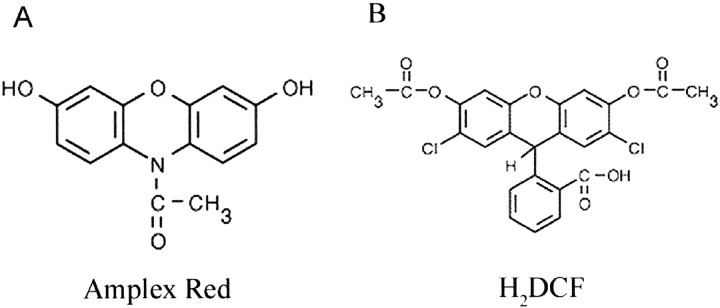
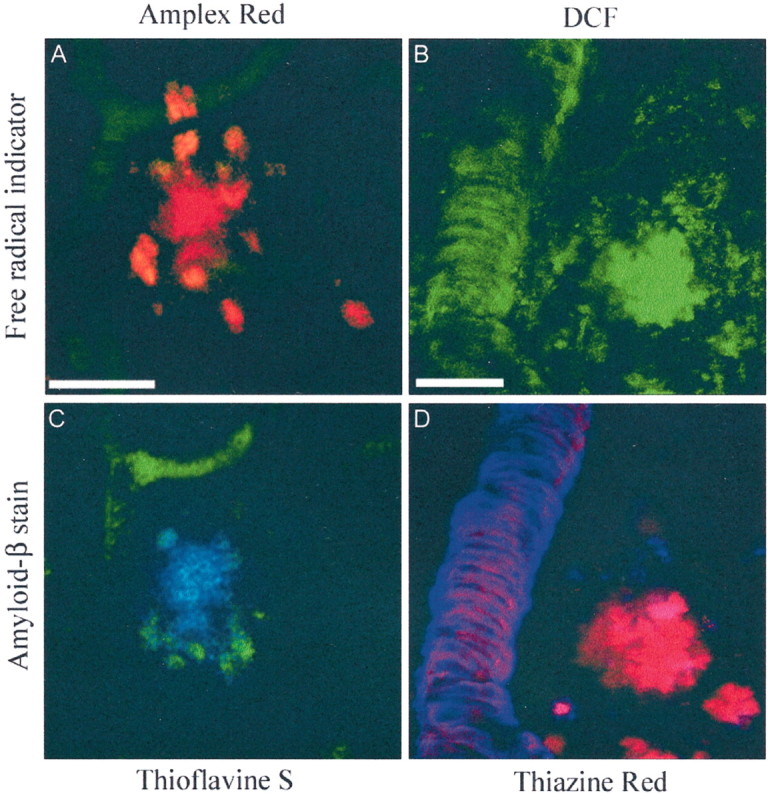
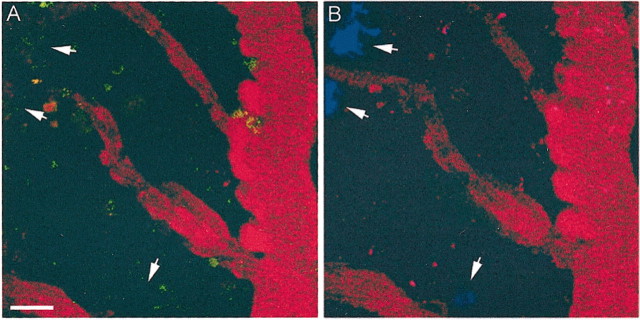
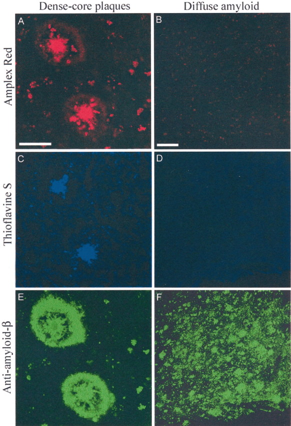
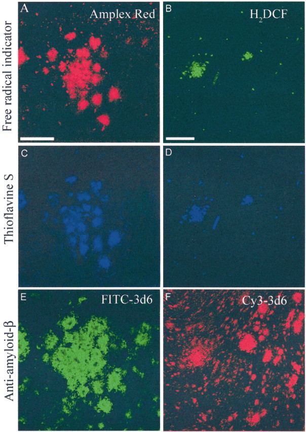
Amyloid-beta, the primary constituent of senile plaques in Alzheimer's disease, is hypothesized to cause neuronal damage and cognitive failure, but the mechanisms are unknown. Using multiphoton imaging, we show a direct association between amyloid-beta deposits and free radical production in vivo in live, transgenic mouse models of Alzheimer's disease and in analogous ex vivo experiments in human Alzheimer tissue. We applied two fluorogenic compounds, which become fluorescent only after oxidation, before imaging with a near infrared laser. We observed fluorescence associated with dense core plaques, but not diffuse plaques, as determined by subsequent addition of thioflavine S and immunohistochemistry for amyloid-beta. Systemic administration of N-tert-butyl-alpha-phenylnitrone, a free radical spin trap, greatly reduced oxidation of the probes. These data show directly that a subset of amyloid plaques produces free radicals in living, Alzheimer's models and in human Alzheimer tissue. Antioxidant therapy neutralizes these highly reactive molecules and may therefore be of therapeutic value in Alzheimer's disease.


Figures

Fig. 1.
Fig. 1.
Free radical indicators are chemically distinct. The structures and properties of the two fluorogenic indicators of oxidative stress, Amplex Red (A) and H2DCF (B), are unique.
Fig. 2.
Fig. 2.
A subset of amyloid-β plaques oxidizes free radical indicators in vivo. Dense core amyloid-β plaques activate the fluorogenic free radical indicators Amplex Red (A) and H2DCF (B) in vivo in live, PDAPP mouse cortex. Histochemical markers of dense core plaques, thioflavine S (C) and thiazine red (D), respectively, confirm these results. Vessel-associated amyloid angiopathy occasionally activated the probes (B), confirmed here with thiazine red (D). Autofluorescent lipofuscin was observed near plaques in multiple optical channels (A, orange). Fluorescein-containing blood vessels used to map sites for reimaging are shown ingreen in A and C. Scale bars: A, C, 10 μm; B, D, 25 μm.
Fig. 3.
Fig. 3.
PBN can prevent plaque-associated oxidationin vivo. Systemic administration of a free radical spin trap, PBN, prevents oxidation of H2DCF (A) by dense core plaques that are clearly visualized with thioflavine S (B).Arrows indicate the absence of DCF-labeled plaques (A) in the same location that contained thioflavine S-positive plaques (B).Green fluorescence in A is caused by autofluorescent lipofuscin. Blood vessels used as a site map for reimaging are shown in red. Scale bar, 25 μm.
Fig. 4.
Fig. 4.
Dense core plaques produce free radicals ex vivo in human AD tissue. Amplex Red was tested in human AD brain tissue containing only dense core plaques (A, C, E) or only diffuse amyloid (B, D, F). Dense core plaques oxidize Amplex Red (A), but diffuse amyloid-β does not (B). Thioflavine S staining (C, D, respectively) and anti-amyloid-β immunohistochemistry (E, F, respectively), in the same tissue sections confirm that Amplex Red associates specifically with thioflavine S- positive dense core plaques, but not larger areas of diffuse amyloid-β alone. Similar results were seen using H2DCF as the free radical indicator (data not shown). Scale bars: A, C, E, 50 μm; B, D, F, 100 μm.
Fig. 5.
Fig. 5.
Dense core plaques produce free radicals ex vivo in PDAPP mouse tissue. Similar to human AD tissue, dense core plaques also oxidize Amplex Red (A) and H2DCF (B) in PDAPP mouse brain tissue. Thioflavine S labeling (C, D, respectively) and anti-amyloid-β immunohistochemistry (E, F, respectively), in the same tissue sections again reveal that dense core plaques, but not diffuse plaques, oxidize the free radical probes. Scale bars: A, C, E, 50 μm; B, D, F, 100 μm.

Similar articles

Cited by

References

    1. Bacskai BJ, Kajdasz ST, Christie RH, Carter C, Games D, Seubert P, Schenk D, Hyman BT. Imaging of amyloid-beta deposits in brains of living mice permits direct observation of clearance of plaques with immunotherapy. Nat Med. 2001;7:369–372. - PubMed
    1. Bacskai BJ, Kajdasz ST, McLellan ME, Games D, Seubert P, Schenk D, Hyman BT. Non-Fc-mediated mechanisms are involved in clearance of amyloid-beta in vivo by immunotherapy. J Neurosci. 2002;22:7873–7878. - PMC - PubMed
    1. Behl C, Davis J, Cole GM, Schubert D. Vitamin E protects nerve cells from amyloid beta protein toxicity. Biochem Biophys Res Commun. 1992;186:944–950. - PubMed
    1. Behl C, Davis JB, Lesley R, Schubert D. Hydrogen peroxide mediates amyloid beta protein toxicity. Cell. 1994;77:817–827. - PubMed
    1. Bruce AJ, Malfroy B, Baudry M. beta-Amyloid toxicity in organotypic hippocampal cultures: protection by EUK-8, a synthetic catalytic free radical scavenger. Proc Natl Acad Sci USA. 1996;93:2312–2316. - PMC - PubMed

Publication types

MeSH terms