. 2003 Mar 15;23(6):2228-38.
doi: 10.1523/JNEUROSCI.23-06-02228.2003.

Adenylate cyclase 1 as a key actor in the refinement of retinal projection maps

Affiliations

Adenylate cyclase 1 as a key actor in the refinement of retinal projection maps

Anne Ravary et al. J Neurosci. .

Abstract

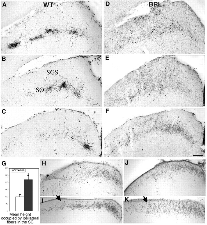
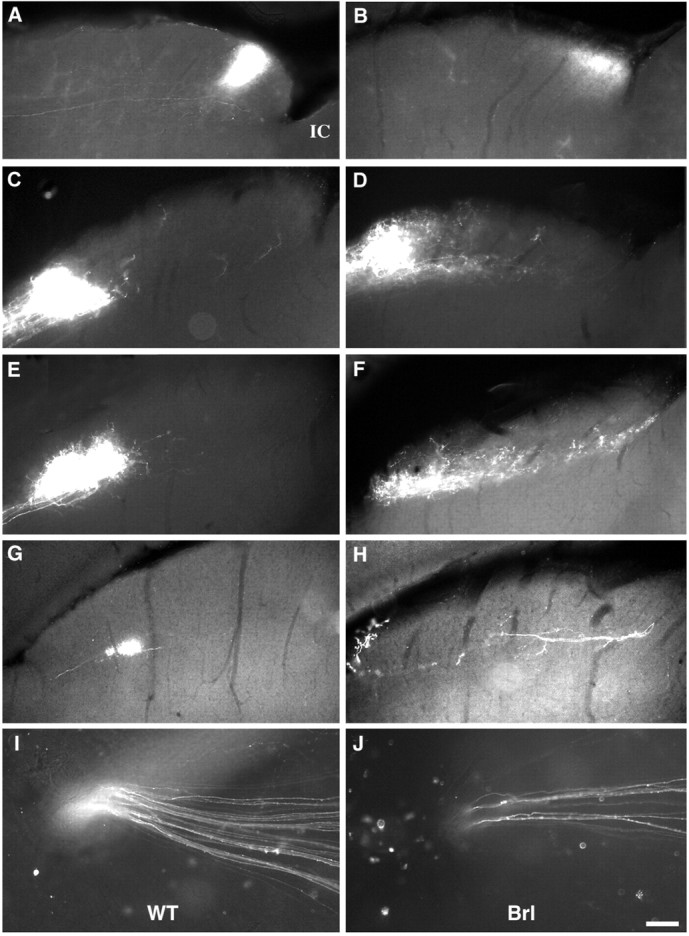
cAMP occupies a strategic position to control neuronal responses to a large variety of developmental cues. We have analyzed the role of calcium-stimulated adenylate cyclase 1 (AC1) in the development of retinal topographic maps. AC1 is expressed in retinal ganglion cells (RGCs) from embryonic day 15 to adulthood with a peak during the first postnatal week. At that time, the other calcium-stimulated AC, AC8, is expressed in the superior colliculus (SC) but not in the RGCs. In mice of the barrelless strain, which carry an inactivating mutation of the AC1 gene, calcium-stimulated AC activity is reduced by 40-60% in the SC and retina. RGC projection maps were analyzed with a variety of anterograde and retrograde tracers. After an initially normal development until postnatal day 3, retinal fibers from the ipsilateral and contralateral eye fail to segregate into eye-specific domains in the lateral geniculate nucleus and the SC. Topographic defects in the fine tuning of the retinotectal and retinogeniculate maps are also observed with abnormalities in the confinement of the retinal axon arbors in the anteroposterior and mediolateral dimensions. This is attributable to the lack of elimination of misplaced axon collaterals and to the maintenance of a transient ipsilateral projection. These results establish an essential role of AC1 in the fine patterning of the retinal map. Calcium-modulated cAMP production in the RGCs could constitute an important link between activity-dependent changes and the anatomical restructuring of the retinal terminal arbors within central targets.


Figures

Fig. 1.
Fig. 1.
Localization of AC1 and AC8 mRNA in the developing visual system. AC1 (left) and AC8 (right) mRNAs were revealed with digoxigenin-labeled probes (A–E) or with radiolabeled probes (F) on cryostat sections from P3 pups (WT mice). A, B, In the retina, strong homogenous AC1 expression (A) is detected in all the RGCs as shown at a higher magnification (A′) and appears to be limited to this neuronal population. With the AC8 probe, no specific signal is detected (B); the diffuse gray stain is identical to that obtained with control sense probes. C, D, In the thalamus, AC1 gene expression (C) was detected in the dLGN, the ventrobasal thalamic nucleus (VB), and the reticulans (RT) but there is no visible AC8 signal. Some AC8 expression is observed in the habenula (Hb) (the sections are in the coronal plane; medial is to theright, and dorsal is at the top).E, A very low level of AC1 is detected in the SC, contrasting with the high expression that is visible in the inferior colliculus (IC). F, Significant AC8 expression is detected in the upper layers of the SC. Scale bar: A–F, 0.2 mm; A′, 0.05 mm.
Fig. 2.
Fig. 2.
AC activity in the developing retinotectal system.A, Calcium-stimulated activity in the SC (black squares) and in the retina (gray circles) during development in the WT mice. The solid curves show the AC activity in the presence of 300 nm calcium, and the dotted curves show the basal AC activity without free calcium. Calcium-stimulated AC activity is higher in the SC than in the retina at all the ages tested.B, Calcium-stimulated activity (in the presence of 300 nm calcium) in WT mice (white bars) andbrl mice (black bars) aged P6 in the SC and the retina. The activity is decreased by 40% in both structures. This was replicated in two independent experiments. C, AC activity in the presence of forskolin (50 μm) in WT and brl mice aged P6 in the SC and the retinas. As shown in two independent experiments, there was no change or only a slight increase in the total level of AC in SC and retinas. AC activity is presented as picomoles of cAMP synthesized per milligram of total protein per minute. Error bars indicate SD among triplicates.
Fig. 3.
Fig. 3.
Abnormal segregation of the ipsilateral and contralateral retinal axons in the dLGN of brl mice. Retinal projections were labeled with HRP injected into one eye in adult (A–D) and P3 (F–G) mice. A, B, In WT mice, the contralateral retinal fibers (A) fill the entire dLGN, leaving a small unlabeled territory in the central region of the dLGN (arrow), whereas the ipsilateral retinal projections (B) are distributed in a dense mediolateral patch. C, D, In the brl mice, the contralateral retinal axons (C) fill the entire dLGN without leaving a gap, whereas the ipsilateral retinal axons (D) are very loosely and widely distributed in the dLGN. E, The fraction of the total dLGN volume that is occupied by the ipsilateral terminals was measured on complete series of sections through the dLGN. The volume occupied by the ipsilateral RGCs is significantly larger in the brl mice (black bars) than in the WT mice (open bars). The mean values and SDs are calculated from five cases of each genotype. *Significant difference (ANOVA, p< 0.05). F, G, Ipsilateral retinal projections to the dLGN in P3 mice showing a similar widespread distribution in both the WT (F) and the brl(G) strains (n = 4 for each genotype). Scale bar, 0.1 mm.
Fig. 4.
Fig. 4.
Abnormal segregation of the ipsilateral and contralateral retinal axons in the dLGN of brl mice. CTB coupled to Alexa 594 was injected into the ipsilateral eye and Alexa 488 was injected into the contralateral eye of adult WT andbrl mice; sections through the midlevel of the dLGN were analyzed. A, C, In WT mice, retinal axon terminals originating in each eye terminate into nonoverlapping domains, and there appears to be an almost complete exclusion of thered- and green-labeled terminals.B, D, in the brl mice, axon terminals originating from both eyes are intertwined over a large portion of the dLGN; red-labeled ipsilateral axons are found in the midst of the contralateral projection zone; and, conversely, contralateral axons are found in the ipsilateral zone, with no clear frontiers between both labels. Scale bar: A, B, 137 μm; C, D, 34 μm.
Fig. 5.
Fig. 5.
Lack of clustering and abnormal laminar distribution of the ipsilateral retinal projection in the SC ofbrl mice. The retinal projections in the SC were HRP-labeled in 5-week-old (A–F) and P3 (H–K) mice of the WT andbrl strains. A–C, In the WT mice, the ipsilateral fibers are clustered in the deep layer of the superior colliculus, the SO, with very few axons entering the upper layer, the SGS. As shown on three coronal sections (spaced by 200 μm) through the SC, four clusters are visible in the rostral SC (A); three clusters are seen at intermediate levels of the SC (B); and only one is present in the caudal SC (C). In thebrl mice, the ipsilateral fibers do not aggregate as clusters at any level of the SC. The ipsilateral retinal fibers have a broader mediolateral extension in the caudal SC (compare F, C) and invade the SGS. G, The mean height of the ipsilateral retinal axons in the SC was quantified and showed a twofold increase in the brl mice (black bar) compared with the controls (white bar). Measures were done as follows. The second, fifth, and ninth sections of the complete rostrocaudal series through the SC were photographed; the area containing the HRP labeling was delimited; and the mean height was calculated. The values are normalized relative to controls. Means and SDs are calculated from five cases for each genotype. *Significant difference (ANOVA, p < 0.05). H–K, At P3, the ipsilateral retinal fibers are not yet clustered in the WT mice (H, I) in either the rostral SC (H) or caudal SC (I) (n = 4 for each genotype). J, K, A similar distribution is noted in rostral (J) and caudal (K) sections through the SC of thebrl mice. However, the retinal fibers have a more diffuse extension in the ventrodorsal plane in the brlmice in comparison with controls (arrow). Scale bar:A–F, 0.1 mm; H–K, 0.09 mm.
Fig. 6.
Fig. 6.
Altered retinotopic projections in the retinotectal system of the brl mice. Small crystals of DiI were placed in the nasal retina (A, B) or the temporal retina (C–J) in WT mice (A, C, E, G) and in brl mice (B, D, F, H). Cases in which only a small amount of RGC axons were labeled (I, J), were selected for analysis. Serial Vibratome sagittal sections (140 μm thick) were made through the SC to measure the extent of the nasal or the temporal retinal projections. In the micrographs of the SC, rostral is to theleft. A, B, Nasal RGCs project in the caudal part of the contralateral SC in both WT mice (A) and brl mice (B). In the brl mice, the projection is much more loosely distributed than in the WT mice and occupies a significantly wider area (see quantifications in Table 2).C–F, Temporal RGCs to the contralateral SC are clustered in the rostral part of the SC, with only a few labeled fibers extending caudally to this patch in WT mice, for which two different cases are illustrated (C, E). In thisbrl mouse (D, F), the temporal retinal projection is less dense and extends to occupy the rostral half of the SC in some cases (D) or its entire rostrocaudal extent in other cases (F).G, H, Projections from the temporal retina to the ipsilateral SC are scarce, generally limited to one or two fibers, which form a dense cluster in WT mice (G), or are spread out in the brl, with only a few collateral branches (H). I,J, DiI injections in the retina corresponding to the cases in E and F are shown to illustrate the small number of the labeled RGCs that converge toward the optic disk. Scale bar: A–F, 0.1 mm; G–H, 0.05 mm.
Fig. 7.
Fig. 7.
Lack of focusing of retinotopic projections in the dLGN of the brl mice. Small crystals of DiI were placed in the temporal retina, and the contralateral dLGN was examined on coronal sections in WT mice (A–C) andbrl mice (D–F). Three different cases are illustrated. In all WT cases (A–C), the projection formed by the temporal retinal axons forms a dense patch in the medioventral part of the dLGN. In the brl cases, with similarly sized injections in the retina, the terminal field occupies a larger extent of the dLGN (D, E) and appears to be more scattered (F). Scale bar, 155 μm.
Fig. 8.
Fig. 8.
Distribution of RGCs in the retina of adult WT andbrl mice. A, Semithin thick transverse sections (1 μm thick stained with toluidine blue) through the adult retinas of WT mice (top) and brl mice (bottom). The RGCs layer is noted with anarrow; the outer layer (photoreceptors) is at thetop. In both the WT retinas (top) and thebrl retinas (bottom), the RGCs (arrows) are distributed as a continuous monolayer. Scale bar, 5 μm. B, Distribution of the ipsilateral RGCs. Camera lucida drawings of the flattened retinas of adult WT andbrl mice after fluorogold injections in the ipsilateral optic tract are shown. Most of the ipsilaterally projecting cells are localized in a ventrotemporal crescent (black) covering a similar size in both genotypes. RGCs situated outside this crescent were plotted individually. A fourfold to fivefold increase of these ectopic ipsilateral RGCs was noted in the brl mice (Table 3). OD, Optic disk; N, nasal;T, temporal. Dorsal is at the top.

References

    1. Abdel-Majid RM, Leong WL, Schalkwyk LC, Smallman DS, Wong ST, Storm DR, Fine A, Dobson MJ, Guernsey DL, Neumann PE. Loss of adenylate cyclase I activity disrupts patterning of mouse somatosensory cortex. Nat Genet. 1998;19:289–291. - PubMed
    1. Abrams TW, Karl KA, Kandel ER. Biochemical studies of stimulus convergence during classical conditioning in Aplysia: dual regulation of adenylate cyclase by Ca2+/calmodulin and transmitter. J Neurosci. 1991;11:2655–2665. - PMC - PubMed
    1. Brooks SP, Storey KB. Bound and determined: a computer program for making buffers of defined ion concentrations. Anal Biochem. 1992;201:119–126. - PubMed
    1. Cai D, Qiu J, Cao Z, McAtee M, Bregman BS, Filbin MT. Neuronal cyclic AMP controls the developmental loss in ability of axons to regenerate. J Neurosci. 2001;21:4731–4739. - PMC - PubMed
    1. Cali JJ, Zwaagstra JC, Mons N, Cooper DM, Krupinski J. Type VIII adenylate cyclase: a Ca2+/calmodulin-stimulated enzyme expressed in discrete regions of rat brain. J Biol Chem. 1994;269:12190–12195. - PubMed

Publication types

MeSH terms

LinkOut - more resources