. 2003 Mar 15;23(6):2265-73.
doi: 10.1523/JNEUROSCI.23-06-02265.2003.

Proteolipid protein gene mutation induces altered ventilatory response to hypoxia in the myelin-deficient rat

Affiliations

Proteolipid protein gene mutation induces altered ventilatory response to hypoxia in the myelin-deficient rat

Martha J Miller et al. J Neurosci. .

Abstract

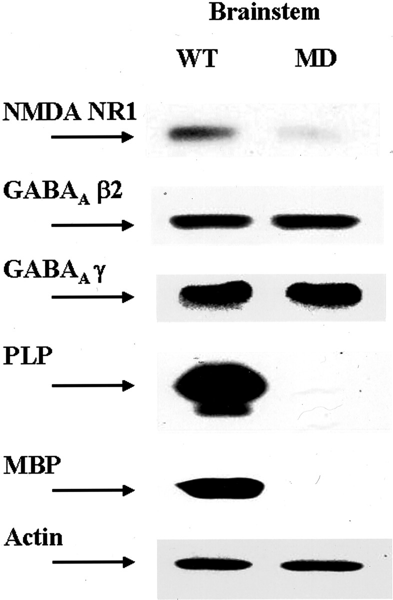
Pelizaeus Merzbacher disease is an X-linked dysmyelinating disorder of the CNS, resulting from mutations in the proteolipid protein (PLP) gene. An animal model for this disorder, the myelin-deficient (MD) rat, carries a point mutation in the PLP gene and exhibits a phenotype similar to the fatal, connatal disease, including extensive dysmyelination, tremors, ataxia, and death at approximately postnatal day 21 (P21). We postulated that early death might result from disruption of myelinated neural pathways in the caudal brainstem and altered ventilatory response to oxygen deprivation or hypercapnic stimulus. Using barometric plethysmography to measure respiratory function, we found that the MD rat develops lethal hypoxic depression of breathing at P21, but hypercapnic ventilatory response is normal. Histologic examination of the caudal brainstem in the MD rat at this age showed extensive dysmyelination and downregulation of NMDA and to a lesser extent GABA(A) receptors on neurons in the nucleus tractus solitarius, hypoglossal nucleus, and dorsal motor nucleus of the vagus. Unexpectedly, immunoreactive PLP/DM20 was detected in neurons in the caudal brainstem. Not all biosynthetic functions and structural elements were altered in these neurons, because phosphorylated and nonphosphorylated neurofilament and choline acetyltransferase expression were comparable between MD and wild-type rats. These findings suggest that PLP is expressed in neurons in the developing brainstem and that PLP gene mutation can selectively disrupt central processing of afferent neural input from peripheral chemoreceptors, leaving the central chemosensory system for hypercapnia intact.


Figures

Fig. 1.
Fig. 1.
Ventilatory response to inhalation of 8% O2 or 10% CO2 in the wild-type (n = 19) and MD (n = 18) rat. MD affected males (open triangles) and normal male littermates (filled squares) exhibited similar responses to inhalation of 8% O2 for 5 min (P14,A; P18, B). At P21–P24, MD rats exhibited significant depression of ventilation in response to 8% O2 compared with wild-type males (C) (*p < 0.05). At all ages, the ventilatory response to inhalation of 10% CO2 for 10 min. was not different in the MD and wild-type rats (P14, D; P18, E; P21–P24,F).
Fig. 2.
Fig. 2.
Cells stained with cresyl violet were evenly distributed at the level of the area postrema in the caudal brainstem in P21 wild-type rats (A) and MD rats (B). Scale bar, 100 μm.
Fig. 3.
Fig. 3.
NeuN expression in the MD and WT rat at P21. Neurons were stained throughout the caudal brainstem at the level of the area postrema in the wild-type rat (A) and MD rat (B). There appeared to be reduced staining for NeuN in the nucleus tractus solitarius and the hypoglossal nucleus (B, D), which was not significant when quantified (Table 1). Boxed areas in Aand B are enlarged in C andD. Scale bars: A, B, 100 μm; C, D, 50 μm.
Fig. 4.
Fig. 4.
Immunoreactivity for nonphosphorylated neurofilament in the caudal brainstem was distributed similarly in wild-type (A) and MD (B) rats at P21. Boxed areas in A andB are enlarged in C and D. Scale bars: A, B, 100 μm;C, D, 50 μm.
Fig. 5.
Fig. 5.
Expression of PLP in the wild-type and MD rat at P21. In the caudal brainstem, immunostaining for PLP was greatly diminished in the MD rat (B) compared with the wild type (A). Furthermore, neurons immunoreactive for PLP were present in the hypoglossal nucleus (B). When sections from the wild-type rat were double labeled for NeuN (red) and PLP/DM20 (green), no cells immunoreactive for both were observed (C). However, in the MD rat, cells were found that were double labeled for NeuN and PLP/DM20 (yellow) (D). Boxed areas in A and B represent comparable areas with the sites imaged in C andD. Scale bars: A, B, 100 μm; C, D, 33 μm.
Fig. 6.
Fig. 6.
In transgenic mice (P12) expressing thePLP promoter driving expression of EGFP (B), neurons (yellow) were found in the nucleus tractus solitarius that were immunoreactive for NeuN (red) and also expressed EGFP-PLP(green). Nontransgenic mice expressed no autofluorescence in neurons in the NTS (A). Scale bars: A, B, 33 μm.
Fig. 7.
Fig. 7.
NMDA NR1-immunoreactive neurons in the WT and MD rat at P21. In the caudal medulla, at the level of the area postrema, NMDA NR1-reactive neurons were observed in the nucleus tractus solitarius, hypoglossal nucleus, and dorsal motor nucleus of the vagus (A). In contrast, in the MD rat, neurons immunoreactive for NMDA NR1 were strikingly diminished in these areas (B). Boxed areas in A and B are enlarged inC–F: top box, C,D; bottom box, E,F. Scale bars: A, B, 100 μm; C–F, 50 μm.
Fig. 8.
Fig. 8.
GABAA β2 subunit immunoreactivity in the WT and MD rat at P21. In the caudal brainstem, at the level of the area postrema, GABAA β2 subunit-immunoreactive neurons were present in the nucleus tractus solitarius, dorsal motor nucleus of the vagus, and hypoglossal nucleus (A). Immunoreactivity for this receptor was diminished in the MD rat at P21 (B). Scale bars: A,B, 100 μm.
Fig. 9.
Fig. 9.
Immunoreactivity for ChAT was similar in the WT (A) and MD (B) rat in the DMV and hypoglossal nuclei. Scale bars: A,B, 100 μm.
Fig. 10.
Fig. 10.
Expression of PLP/DM20, MBP, NMDA NR1, and GABAA β2 and γ subunits assayed by immunoblot in tissue from the brainstem in the wild-type and MD rat at P21. Actin is present as a loading control. In the MD rat, there was downregulation of NMDA NR1, PLP/DM20, and MBP.

Similar articles

Cited by

References

    1. Ang RC, Hoop B, Kazemi H. Role of glutamate as the central neurotransmitter in the hypoxic ventilatory response. J Appl Physiol. 1992;72:1480–1487. - PubMed
    1. Boison D, Stoffel W. A point mutation in exon 3 (A-C, Thr 75-Pro) of the myelin proteolipid protein causes dysmyelination and oligodendrocyte death. EMBO J. 1989;8:3295–3302. - PMC - PubMed
    1. Boulloche J, Aicardi J. Pelizaeus-Merzbacher disease: clinical and nosological study. J Child Neurol. 1986;1:233–239. - PubMed
    1. Campagnoni CW, Garbay B, Micevych P, Pribyl T, Kampf K, Handley VW, Campagnoni AT. DM20 mRNA splice products of the myelin proteolipid protein gene are expressed in the murine heart. J Neurosci Res. 1992;33:148–155. - PubMed
    1. Ciriello J, Hrycyshyn AW, Calaresu FR. Horseradish peroxidase study of brain stem projections of carotid sinus and aortic depressor nerves in the cat. J Auton Nerv Syst. 1981;4:43–61. - PubMed

Publication types

MeSH terms