. 2003 Mar 15;23(6):2371-82.
doi: 10.1523/JNEUROSCI.23-06-02371.2003.

Regulation of extracellular signal-regulated kinase by cannabinoids in hippocampus

Affiliations

Regulation of extracellular signal-regulated kinase by cannabinoids in hippocampus

Pascal Derkinderen et al. J Neurosci. .

Abstract

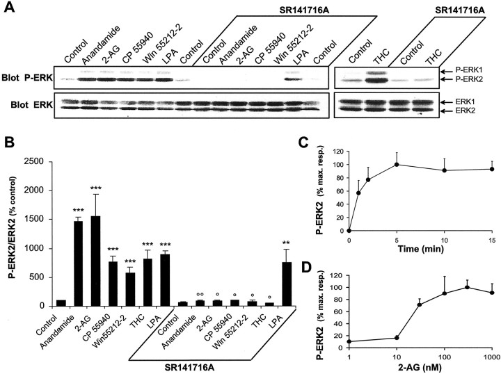
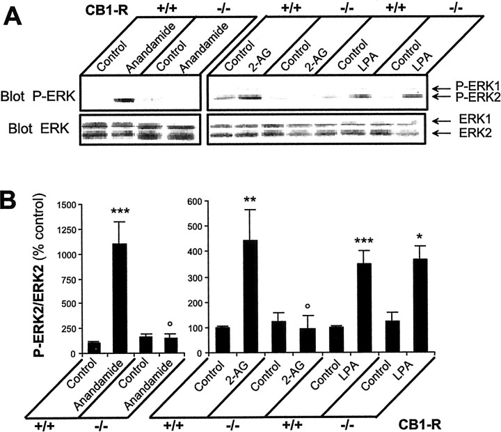
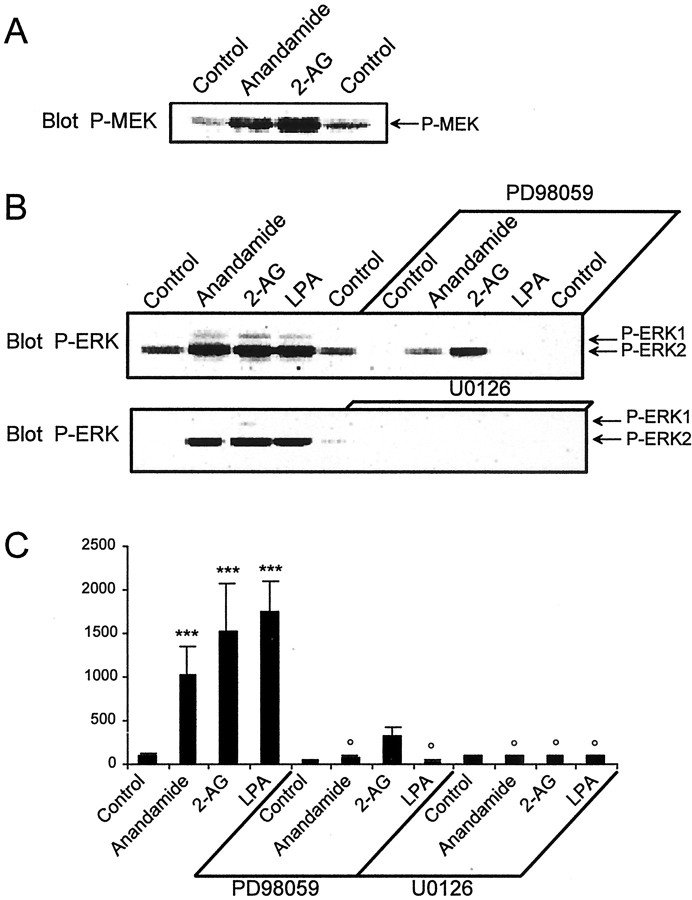
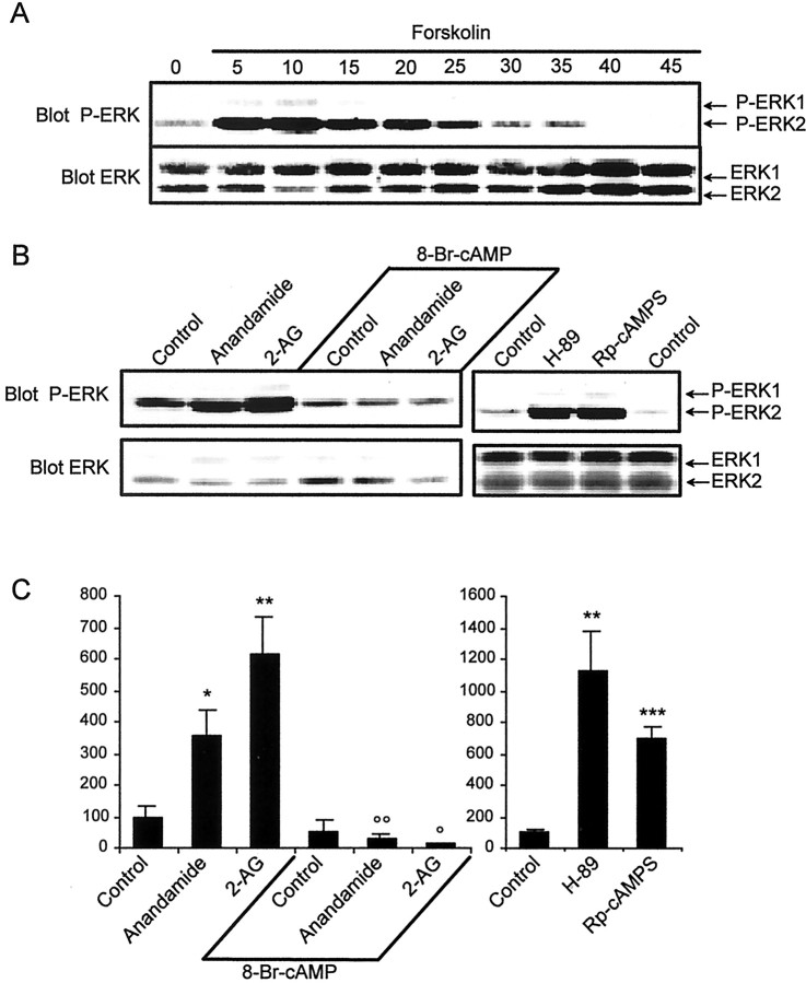
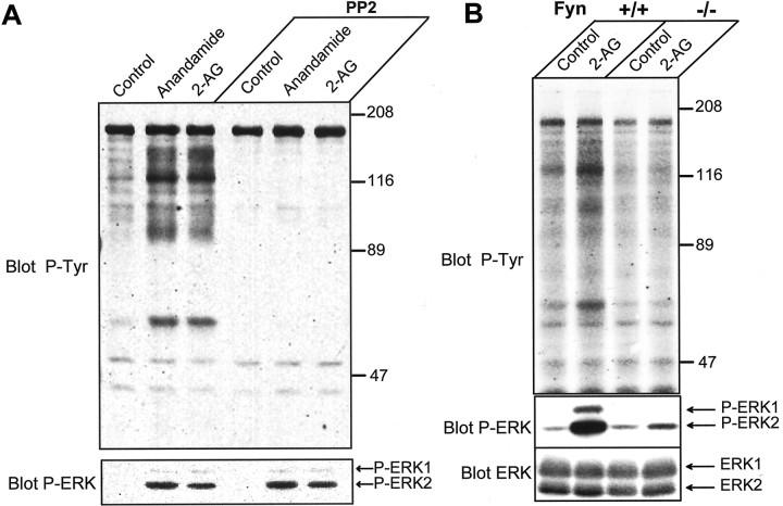
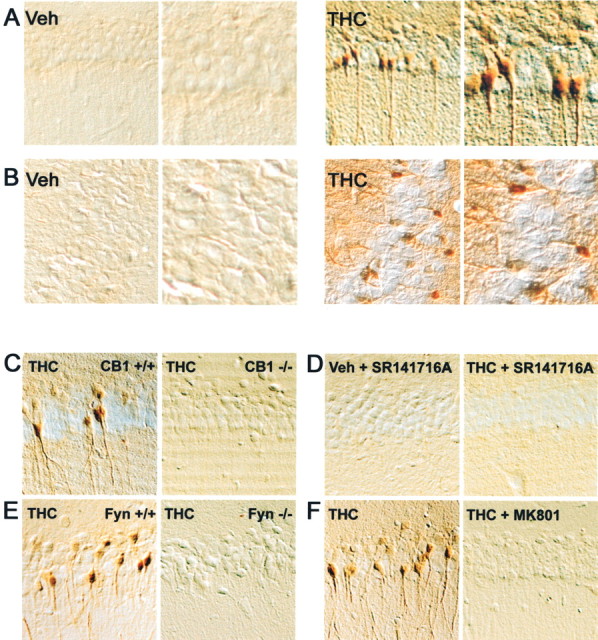
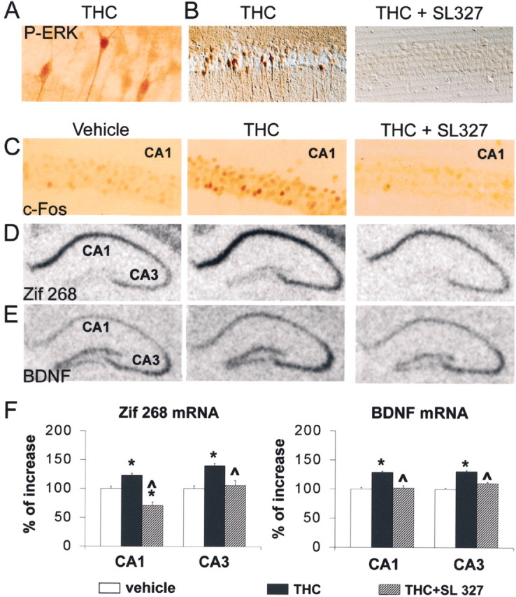
Endocannabinoids form a novel class of intercellular messengers, the functions of which include retrograde signaling in the brain and mediation or modulation of several types of synaptic plasticity. Yet, the signaling mechanisms and long-term effects of the stimulation of CB1 cannabinoid receptors (CB1-R) are poorly understood. We show that anandamide, 2-arachidonoyl-glycerol, and Delta9-tetrahydrocannabinol (THC) activated extracellular signal-regulated kinase (ERK) in hippocampal slices. In living mice, THC activated ERK in hippocampal neurons and induced its accumulation in the nuclei of pyramidal cells in CA1 and CA3. Both effects were attributable to stimulation of CB1-R and activation of MAP kinase/ERK kinase (MEK). In hippocampal slices, the stimulation of ERK was independent of phosphatidyl-inositol-3-kinase but was regulated by cAMP. The endocannabinoid-induced stimulation of ERK was lost in Fyn knock-out mice, in slices and in vivo, although it was insensitive to inhibitors of Src-family tyrosine kinases in vitro, suggesting a noncatalytic role of Fyn. Finally, the effects of cannabinoids on ERK activation were dependent on the activity of glutamate NMDA receptors in vivo, but not in hippocampal slices, indicating the existence of several pathways linking CB1-R to the ERK cascade. In vivo THC induced the expression of immediate-early genes products (c-Fos protein, Zif268, and BDNF mRNAs), and this induction was prevented by an inhibitor of MEK. The strong potential of cannabinoids for inducing long-term alterations in hippocampal neurons through the activation of the ERK pathway may be important for the physiological control of synaptic plasticity and for the general effects of THC in the context of drug abuse.


Figures

Fig. 1.
Fig. 1.
Cannabinoid agonists stimulate ERK phosphorylation in rat hippocampal slices. A, Rat hippocampal slices were incubated at 35°C, as described in Materials and Methods, for 50 min before the addition of vehicle (Control), 1 μm anandamide, 1 μm 2-AG, 1 μm CP 55940, 100 μm WIN 55212–2, 0.2 μm LPA, or 0.1 μm Δ9-THC for 5 min, in the absence or in the presence of 100 μm SR 141716A applied 30 min before. Slices were homogenized in SDS; 60 μg of protein per sample were subjected to immunoblot analysis using antibodies specific for the dually phosphorylated (active) forms of ERK1 and ERK2 (Blot P-ERK). After stripping, the membranes were reprobed with anti-ERK (Blot ERK) antibodies. B, For quantification the optical densities of P-ERK2-immunoreactive bands were measured, normalized to the optical densities of total ERK2 in the same samples, and expressed as percentages of controls. Data correspond to means ± SEM. Statistical analysis was done with ANOVA (F(13,24) = 22.8; p< 0.0001) followed by t test (treated vs control: ***p < 0.001, **p < 0.01; treated in the presence of SR141716A vs in its absence: °° p < 0.01, ° p < 0.05). C, D, Quantification of the effects of 2-AG on ERK2 active form: time course (drug concentration 1 μm) (C); concentration–response curve (treatment for 5 min) (D). Immunoreactivity was quantified by scanning densitometry using NIH image 1.62 software. Values are means ± SEM of four to eight independent experiments and are expressed as percentages of the maximal increase above unstimulated control values.
Fig. 2.
Fig. 2.
The regulation of ERKs by endocannabinoids is absent in hippocampal slices from CB1-R knock-out mice.A, Hippocampal slices from wild-type (CB1-R +/+) and CB1-R knock-out (CB1-R −/−) mice were incubated at 35°C, as described in Materials and Methods, for 50 min before the addition of vehicle (Control), 1 μm anandamide, 1 μm 2-AG, or 0.2 μm LPA for 5 min. ERK phosphorylation was assayed as described in the legend to Figure 1. B, Quantification of the results for P-ERK2 as described in the legend to Figure 1. Data correspond to means ± SEM. Statistical analysis was done with ANOVA (anandamide: F(3,20) = 40.2,p < 0.0001; 2-AG and LPA:F(5,24) = 8.2, p < 0.0001) followed by t test (treated vs control: ***p < 0.001, **p < 0.01, *p < 0.05; treated in knock-out vs wild type: ° p < 0.05).
Fig. 3.
Fig. 3.
Role of MEK in the effects of endocannabinoids in rat hippocampal slices. A, Rat hippocampal slices were incubated as described in Materials and Methods, for 50 min before the addition of vehicle (Control), 1 μmanandamide, or 1 μm 2-AG for 5 min. Homogenates (60 μg of protein per sample) were analyzed by immunoblotting with antibodies specific for the phosphorylated forms of MEK1/2. Quantification of P-MEK immunoreactivity (mean ± SEM): controls 100 ± 8, anandamide 857 ± 29, 2-AG 514 ± 57 (F(2,5) = 226, p < 0.001; t test treated vs control: p< 0.0001). B, Slices were treated with the same compounds as in A, or with 0.2 μm LPA, in the absence or in the presence of 50 μm PD98059 or 30 μm U0126, two MEK inhibitors. Homogenates were analyzed for active dually phosphorylated ERK by immunoblotting.C, Quantification of the results for P-ERK2 as described in the legend to Figure 1. Data correspond to mean ± SEM. Statistical analysis was done with ANOVA (F(11,45 = 7.4; p< 0.0001) followed by t test (treated vs control: ***p < 0.001; treated in the presence of MEK inhibitor vs in its absence: ° p < 0.05).
Fig. 4.
Fig. 4.
Role of cAMP in the effects of endocannabinoids on ERK phosphorylation. A, Rat hippocampal slices were incubated with forskolin (50 μm) for the indicated period of time. Homogenates were analyzed for active dually phosphorylated ERK by immunoblotting. B, Rat hippocampal slices were incubated as described in Materials and Methods, in the presence or in the absence of 4 mm 8-Br-cAMP for 45 min before the addition of vehicle (Control), 1 μmanandamide, or 2-AG for 5 min. In other experiments, slices were incubated in the presence of the PKA inhibitors H-89 (100 μm) or Rp-cAMPS (1 mm) for 20 min. Homogenates were analyzed for active dually phosphorylated ERK by immunoblotting as described in the legend to Figure 1.C, Quantification of the results for P-ERK2 as described in the legend to Figure 1. Data correspond to mean ± SEM. Statistical analysis was done with ANOVA (8-Br-cAMP:F(5,18) = 14.5; H89 and RpcAMPs:F(2,7) = 16.1; p < 0.01) followed by t test (treated vs control: ***p < 0.001, **p < 0.01, *p < 0.05; treated in the presence of 8-Br-cAMP vs in its absence: °° p < 0.01, ° p < 0.05).
Fig. 5.
Fig. 5.
Role of Src-family kinases in the effects of endocannabinoids in rat hippocampal slices. A, Rat hippocampal slices were incubated as described in Materials and Methods, in the presence or absence of 5 μm PP2 for 45 min before the addition of vehicle (Control), 1 μm anandamide, or 2-AG for 5 min. Homogenates were analyzed by immunoblotting with antibodies specific for anti-phospho-tyrosine (Blot P-Tyr), active dually phosphorylated ERK (Blot P-ERK), or total ERK (Blot ERK). The optical densities of P-ERK2 were as follows: in the absence of PP2: control 100 ± 43, anandamide 767 ± 231, and 2-AG 413 ± 92; in the presence of PP2: control 89 ± 28, anandamide 1358 ± 280, and 2-AG 861 ± 191 (F(5,14) = 7.1,p < 0.001; followed by t test,p < 0.05 for endocannabinoid-treated vs control in both groups). B, Hippocampal slices from wild-type (Fyn +/+) and Fyn knock-out (Fyn −/−) mice were incubated for 50 min before the addition of either vehicle (Control) or 1 μm2-AG for 5 min. The optical densities of P-ERK2 were as follows: in Fyn +/+ slices: control 100 ± 8, 2-AG 1198 ± 351; in Fyn −/− slices: control 115 ± 23, 2-AG 108 ± 63 (F(3,8) = 9.3, p < 0.01; followed by t test, p < 0.05, 2-AG vs control in wild-type slices, and p < 0.05, 2-AG in knock-out vs control slices).
Fig. 6.
Fig. 6.
THC activates ERK in hippocampus in vivo by stimulating CB1 receptors. Mice were injected with vehicle (Veh) or THC (1 mg/kg, i.p.) 10 min before they were killed. Active ERK1 and ERK2 were detected by peroxidase immunocytochemistry in the hippocampus, using antibodies against the doubly phosphorylated protein. Results in CA1 (A) and CA3 (B) regions are shown. The right panel corresponds to a higher magnification of the THC-treated sections. In THC-treated mice, immunoreactive cells are mostly present in the pyramidal cell layer of CA1 and CA3. A strong labeling is visible in the cytoplasm (including the dendrites) and the nucleus.C, THC failed to activate ERK in CA1 of CB1 −/− mice, whereas it was active in CB1 +/+ matched controls. D, In the presence of the CB1-R antagonist, SR141716A (3 mg/kg) injected alone (Veh + SR141716A), or 15 min before THC injection (THC + SR141716A), no activation of ERK was observed in CA1. E, THC failed to activate ERK in CA1 of Fyn −/− mice, whereas its effects were present in Fyn +/+ matched controls.F, Stimulation of ERK phosphorylation by THC was abolished by the NMDA receptor antagonist MK801 (0.1 mg/kg), injected 15 min before THC. The results obtained in CB1 −/−, SR141716A-treated, Fyn −/−, and MK801-treated mice in CA3 (data not shown) were similar to those illustrated here in CA1.
Fig. 7.
Fig. 7.
ERK-dependent induction of immediate-early genes by THC in hippocampus in vivo. A, Mice were injected with 1 mg/kg THC 10 min before they were killed, as in Figure 6A, High magnification of a peroxidase-labeled section shows the nuclear staining for dually phosphorylated ERK. B, The effects of THC (middle panel) were prevented in the CA1 region of mice injected with SL327 (100 mg/kg) 60 min before THC (right panel). Results shown are representative of four to six animals for each group. C, c-Fos immunoreactivity (peroxidase reaction) was examined in CA1 of mice injected with either vehicle (Veh) or 1 mg/kg THC 60 min before they were killed. SL327 (100 mg/kg) was injected 60 min before THC.D, Zif268 mRNA expression was analyzed by in situ hybridization in mouse hippocampus 1 hr after injection of vehicle, THC (1 mg/kg, i.p.), or THC and SL327 (100 mg/kg, 60 min before THC). Note the increased hybridization signals in the CA1 and CA3 regions. E, BDNF mRNA expression was analyzed byin situ hybridization in mouse hippocampus 1 hr after injection of vehicle, THC (1 mg/kg, i.p.), or THC and SL327 (100 mg/kg, 60 min before THC). F, Signals for mRNA hybridization were quantified using an image analyzer for six animals for each treatment. Statistical analyses used one-way ANOVA followed by apost hoc comparison with Newman–Keuls test; *p < 0.001 when comparing THC-treated mice with control mice; ^ p < 0.001 when comparing SL + THC with THC alone (n = 6 mice per group).

Similar articles

Cited by

References

    1. Alessi DR, Cuenda A, Cohen P, Dudley DT, Saltiel AR. PD 098059 is a specific inhibitor of the activation of mitogen-activated protein kinase kinase in vitro and in vivo. J Biol Chem. 1995;270:27489–27494. - PubMed
    1. Al-Hayani A, Wease KN, Ross RA, Pertwee RG, Davies SN. The endogenous cannabinoid anandamide activates vanilloid receptors in the rat hippocampal slice. Neuropharmacology. 2001;41:1000–1005. - PubMed
    1. Atkins CM, Selcher JC, Petraitis JJ, Trzaskos JM, Sweatt JD. The MAPK cascade is required for mammalian associative learning. Nat Neurosci. 1998;1:602–609. - PubMed
    1. Barnett G, Licko V, Thompson T. Behavioral pharmacokinetics of marijuana. Psychopharmacology. 1985;85:51–56. - PubMed
    1. Bidaut-Russell M, Devane WA, Howlett AC. Cannabinoid receptors and modulation of cyclic AMP accumulation in the rat brain. J Neurochem. 1990;55:21–26. - PubMed

Publication types

MeSH terms

Substances

LinkOut - more resources