. 2003 Apr 1;22(7):1497-507.
doi: 10.1093/emboj/cdg144.

The structure of Bcl-w reveals a role for the C-terminal residues in modulating biological activity

Affiliations

The structure of Bcl-w reveals a role for the C-terminal residues in modulating biological activity

Mark G Hinds et al. EMBO J. .

Abstract

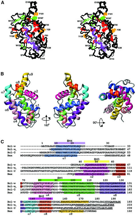
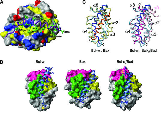
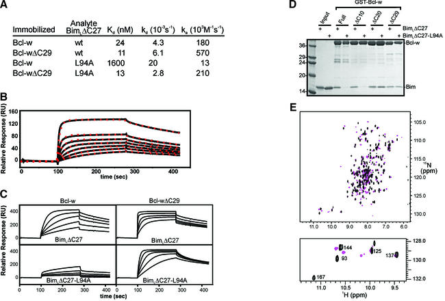
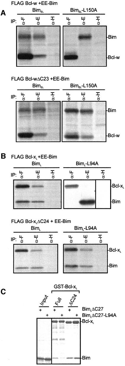
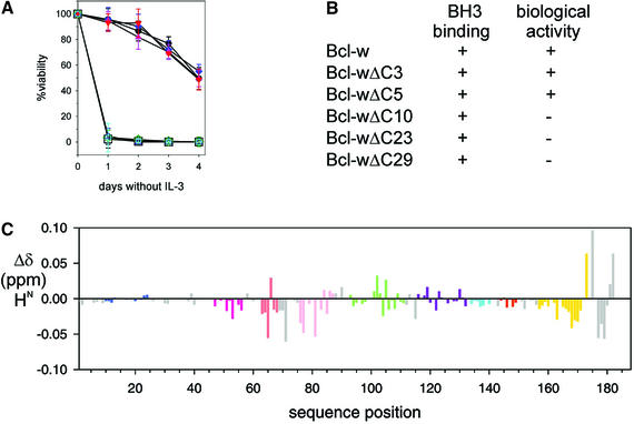
Pro-survival Bcl-2-related proteins, critical regulators of apoptosis, contain a hydrophobic groove targeted for binding by the BH3 domain of the pro-apoptotic BH3-only proteins. The solution structure of the pro-survival protein Bcl-w, presented here, reveals that the binding groove is not freely accessible as predicted by previous structures of pro-survival Bcl-2-like molecules. Unexpectedly, the groove appears to be occluded by the C-terminal residues. Binding and kinetic data suggest that the C-terminal residues of Bcl-w and Bcl-x(L) modulate pro-survival activity by regulating ligand access to the groove. Binding of the BH3-only proteins, critical for cell death initiation, is likely to displace the hydrophobic C-terminal region of Bcl-w and Bcl-x(L). Moreover, Bcl-w does not act only by sequestering the BH3-only proteins. There fore, pro-survival Bcl-2-like molecules probably control the activation of downstream effectors by a mechanism that remains to be elucidated.


Figures

None
Fig. 1. Sequence and structure of Bcl-w. (A) A stereoview of the backbone (N, Cα, C) superposition of the 20 NMR-derived structures of Bcl-wΔC10 (residues 8–183). Aromatic side chains are shown for residues with <25% solvent accessibility in different colors: Trp (green), Phe (red), His (cyan) and Tyr (orange). The region of extended structure at the C-terminus is shown in purple. (B) Ribbon diagram of the structure closest to the mean (residues 8–183). The helices are indicated in different colors and are labeled. The view on the left has the same orientation as (A) while the middle view has been rotated 180° about the vertical axis and the right view 90° about the horizontal axis. (C) Structure-based sequence alignment of Bcl-w, Bcl-xL, Bcl-2 and Bax. The BH domains are indicated by colored bars above the sequence and the helical residues in all the structures are indicated in colors identical to those used in (B). The C-terminal residues, missing from the structures, are underlined.
None
Fig. 2. The hydrophobic binding grooves in Bcl-2 family members. (A) A close-up view of the C-terminal residues of Bcl-w. Residues 8–152 are shown as a surface with the side chains of basic, acidic and hydrophobic residues colored blue, red and yellow, respectively. The C-terminal residues (153–183) are shown as a ribbon (purple) and the side chains of these residues are shown in stick representation (green). (B) Comparison of the hydrophobic binding grooves from Bcl-w, Bax and Bcl-xL. In all three structures residues equivalent to 8–152 in Bcl-w are shown as a surface representation with the BH domains indicated (BH1, green; BH2, pink; BH3, yellow). The residues that lie in the groove (Bcl-w residues 153–181, Bax residues 166–192 and the Bad peptide) are shown as a ribbon (light blue) with the side chains as sticks (blue). The atomic coordinates of Bax (1f16) and the Bcl-xL/Bad (1g5j) peptide complex were obtained from the Protein Data Bank. (C) Comparison of the binding groove in Bcl-w with those in Bax and Bcl-xL. On the left, the ribbon diagram representing Bcl-w (pale blue) is superimposed with Bax (yellow). The C-terminal residues are shown in dark blue (Bcl-w) and orange–yellow (Bax). On the right Bcl-w (pale blue, dark blue) is superimposed with the Bcl-xL (pink):Bad (dark pink) complex. The C-terminus of Bcl-w (C) and the C-termini of Bax and the Bad peptide (C′) are labeled. The structures were superimposed using TOP (Lu, 2000) and the equivalent view is shown for all of them.
None
Fig. 3. Binding properties of Bcl-w proteins. (A) The binding constants were determined using biosensor experiments as described in Materials and methods. (B) Binding of Bcl-w to BimLΔC27 fits a 1:1 model. Samples of serially diluted Bcl-w (2 µM–62.5 nM) were analyzed on a BimLΔC27 sensor surface. The experimental data (solid line) and the suggested fit to a 1:1 Langmuir binding model (red dots) are shown. (C) Interaction kinetics for Bcl-w binding to BimLΔC27. Serial dilutions of Bcl-w or Bcl-wΔC29 were analyzed on parallel sensor surfaces that had been derivatized at comparable densities with either BimLΔC27 or BimLΔC27-L94A. Relative responses of samples between 1 µM and 62.5 nM are shown. (D) GST pull-down assay to assess the binding capacity of Bcl-w proteins. Approximately equivalent amounts of the indicated GST–Bcl-w proteins were mixed with either soluble wild-type BimLΔC27 or soluble BimLΔC27-L94A. The intensity of the Bim band indicated the amount of protein that bound to Bcl-w. Molecular weight standards in kDa are indicated. (E1H-15N-HSQC spectra of 15N Bcl-w in the presence (pink) and absence (black) of a 26 residue Bim-BH3 domain peptide. Inset shows the indole region.
None
Fig. 4. In vivo and in vitro binding properties of Bcl-xL resemble those of Bcl-w. (A) The C-terminal residues of Bcl-w restrict access to the binding groove in vivo. Equivalent 35S-labeled 293T lysates obtained from cells co-expressing FLAG Bcl-w or Bcl-wΔC23, and EE-BimEL or BimEL-L150A, were immunoprecipitated using the anti-FLAG M2 (α-F), anti-EE (α-E) or control anti-HA (α-H) monoclonal antibodies. The immunoprecipitates were fractionated on SDS–PAGE gels. (B) The C-terminal residues of Bcl-xL restrict access to the binding groove in vivo. Co-precipitation experiments similar to those described in (A) using lysates from cells co-expressing FLAG Bcl-xL or Bcl-xLΔC24, and EE-BimL or BimL-L94A. (C) GST pull-down experiment as for Figure 3D, except in this case GST–Bcl-xL proteins were mixed with either soluble wt BimLΔC27 or soluble BimLΔC27-L94A.
None
Fig. 5. Bcl-wΔC10 is functionally inert but is structurally similar to biologically active Bcl-wΔC5. (A) Bcl-w cannot tolerate extensive C-terminal deletions. The viability of parental FDC-P1 cells (black squares) or representative clones expressing different Bcl-w constructs [Bcl-w (black circles); Bcl-w(A128E) (blue); Bcl-wΔC3 (pink); Bcl-wΔC5 (red); Bcl-wΔC10 (green); Bcl-wΔC23 (blue); Bcl-wΔC29 (cyan)] deprived of IL-3 were determined by PI-staining analyzed flow cytometrically. Data shown are means ± 1 SD of at least three experiments. (B) Summary of the binding properties and biological activity of full-length or C-terminal truncated mutants of Bcl-w. (C) Comparison of the 2D 1H-15N-HSQC spectra for Bcl-wΔC10 and Bcl-wΔC5. Backbone amide proton chemical shift differences plotted for residues in 15N-labeled Bcl-wΔC10 relative to those for Bcl-wΔC5 are indicated. Colors for the helices correspond to those used in Figure 1.

References

    1. Aritomi M., Kunishima,N., Inohara,N., Ishibashi,Y., Ohta,S. and Morikawa,K. (1997) Crystal structure of Rat Bcl-xL. J. Biol. Chem., 272, 27886–27892. - PubMed
    1. Bartels C., Xia,T.H., Billeter,M., Güntert,P. and Wüthrich,K. (1995) The program XEASY for computer-supported nmr spectral-analysis of biological macromolecules. J. Biomol. NMR, 6, 1–10. - PubMed
    1. Borner C., Martinou,I., Mattmann,C., Irmler,M., Schaerer,E., Martinou,J.C. and Tschopp,J. (1994) The protein Bcl-2α does not require membrane attachment, but two conserved domains to suppress apoptosis. J. Cell Biol., 126, 1059–1068. - PMC - PubMed
    1. Brünger A.T. et al. (1998) Crystallography and NMR system: a new software suite for macromolecular structure determination. Acta Crystallogr. D., 54, 905–921. - PubMed
    1. Chen-Levy Z., Nourse,J. and Cleary,M.L. (1989) The bcl-2 candidate proto-oncogene product is a 24-kilodalton integral-membrane protein highly expressed in lymphoid cell lines and lymphomas carrying the t(14;18) translocation. Mol. Cell. Biol., 9, 701–710. - PMC - PubMed

Publication types

MeSH terms