. 2003 Apr 1;22(7):1567-78.
doi: 10.1093/emboj/cdg157.

p53 activates ICAM-1 (CD54) expression in an NF-kappaB-independent manner

Affiliations

p53 activates ICAM-1 (CD54) expression in an NF-kappaB-independent manner

Vassilis G Gorgoulis et al. EMBO J. .

Abstract

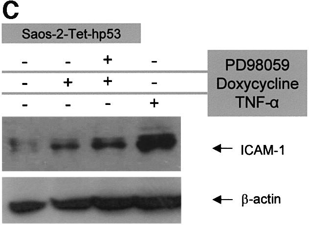
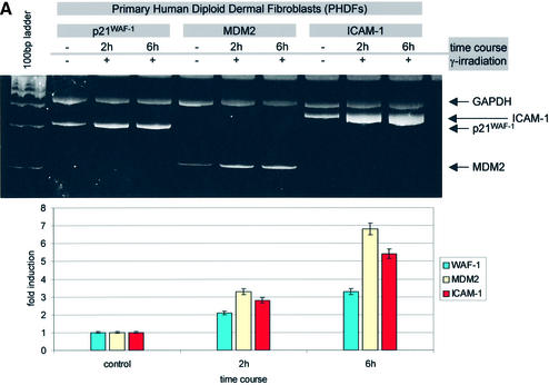
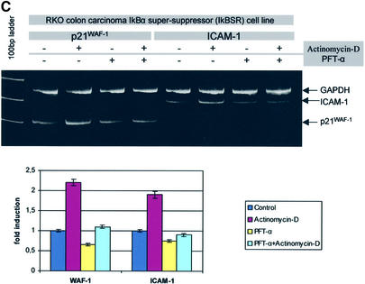
Intercellular adhesion molecule-1 (ICAM-1) is a crucial receptor in the cell-cell interaction, a process central to the reaction to all forms of injury. Its expression is upregulated in response to a variety of inflammatory/immune mediators, including cellular stresses. The NF-kappaB signalling pathway is known to be important for activation of ICAM-1 transcription. Here we demonstrate that ICAM-1 induction represents a new cellular response to p53 activation and that NF-kappaB inhibition does not prevent the effect of p53 on ICAM-1 expression after DNA damage. Induction of ICAM-1 is abolished after treatment with the specific p53 inhibitor pifithrin-alpha and is abrogated in p53-deficient cell lines. Furthermore, we map two functional p53-responsive elements to the introns of the ICAM-1 gene, and show that they confer inducibility to p53 in a fashion similar to other p53 target genes. These results support an NF-kappaB-independent role for p53 in ICAM-1 regulation that may link p53 to ICAM-1 function in various physiological and pathological settings.


Figures

None
Fig. 1. Artificially expressed wt p53 induces ICAM-1 in an NF-κB-independent manner. (A) Treatment of Saos-2-Tet-hp53 cells with the tetracycline analogue Dox showed a 3-fold increase in the ICAM-1 mRNA levels (assessed by comparative multiplex RT–PCR), that remained elevated after administration of the MEK1 inhibitor PD98059, which blocks the Raf/MAPK/pp90rsk/NF-κB pathway (Ryan et al., 2000). The mRNA levels of ICAM-1 in the parental Saos-2 cells were not affected by Dox (not shown). (B) Expression of ICAM-1 (green fluorescence signal) in Saos-2-Tet-hp53 cells under basal conditions (a) and overexpression after Dox (b) and Dox + PD98059 treatment (c). Counterstain with DAPI (intensity normalization was based on DAPI staining). (C) Western immunoblot analysis of ICAM-1 in Saos-2-Tet-hp53 cells after Dox, Dox + PD98059 and TNF-α treatment.
None
Fig. 2. (A) Irradiation-activated p53 in PHDFs induces a 2- and 3.5-fold increase in ICAM-1 mRNA levels, which closely resembles that of the p53 target genes p21WAF-1/CIP-1 and MDM2. (B) Pre-treatment of irradiated PHDFs with the specific p53 inhibitor PFT-α reduces p21WAF-1/CIP-1 and ICAM-1 expression to baseline levels.
None
Fig. 3. Wt p53 and not NF-κB activity is necessary for DNA damage-induced ICAM-1 expression (irradiation or actinomycin D treatment). (A) Irradiation-induced p53 (upper right inset blot) suppresses TNF-α-triggered NF-κB activity. (BICAM-1, p21WAF-1/CIP-1 and MDM2 are not induced in a p53-null environment (Saos-2) following γ-irradiation, as determined by the target/GAPDH ratio which was equal in pre- and post-irradiation measurements. (C) Low dose of the DNA-damaging agent actinomycin D-induced p53 activates p21WAF-1/CIP-1 and ICAM-1 mRNA expression in the NF-κB-inactive environment of the RKO-IκBαSR cells, which falls to baseline levels after treatment with PFT-α.
None
Fig. 4. Genomic structure of the ICAM-1 gene region containing the putative p53REs (contig accession No. AC011511). The ICAM-1 genomic region was found after pairwise homology search (pairwise nucleotide BLAST) of genomic database deposits with the ICAM-1 mRNA sequence (accession No. J03132). The p53REs resembling to a greater extent the classical p53 consensus RRRCWWGYYY (p53CON) (el-Deiry et al., 1992) are marked in red. Additional p53 half-binding sites that match the criteria set by Bourdon et al. (1997) are marked in blue. The mismatches from p53CON are marked in yellow.
None
Fig. 5. In vitro characterization of the p53-binding sites within the intronic sequences of the ICAM-1 gene. (A) EMSA showing specific binding of in vitro produced wt p53 to the identified ICAM-1p53REs (A1, B1, C; el-Deiry consensus). Left panel (A1 probe): lane 1, no retarded ICAM-1p53RE-A1 band in the presence of plain rabbit reticulocyte lysate (RR Lysate); lane 2, in vitro-translated wt p53 protein binds to the labelled specific element in the presence of monoclonal antibody (Ab) PAb421, generating a retarded species; lanes 3 and 4, 50-fold molar excess of unlabelled ICAM-1p53RE-A1 completely abolishes the retarded species, whereas the same amount of mtICAM-1p53RE-A1 [harbouring a mutation at position 4(C) and 7(G) of the consensus] does not affect its formation, demonstrating the specificity of binding; lane 5, in vitro-translated mt p53 protein (V173E) fails to bind the ICAM-1p53RE-A1 element; lane 6, addition of the anti-p53 Ab DO-1, in the presence of PAb421, ‘super-shifted’ the retarded species, verifying the presence of p53 in the DNA–protein complex. Middle panel (B1 probe): lane 1, no retarded ICAM-1p53RE-B1 band in the presence of RR lysate; lane 2, in vitro-translated wt p53 protein binds to the labelled specific element in the presence of monoclonal Ab PAb421, generating a retarded species; lanes 3 and 4, 50-fold molar excess of unlabelled ICAM-1p53RE-B1 completely abolishes the retarded species, whereas the same amount of mtICAM-1p53RE-B1 [harbouring a mutation at position 4(C) and 7(G) of the consensus] does not affect its formation; lane 5, in vitro-translated mt p53 protein (V173E) fails to bind the ICAM-1p53RE-B1 element; lane 6, addition of the anti-p53 Ab DO-1, in the presence of PAb421, ‘super-shifted’ the retarded species. Right panel (C probe): lane 1, in vitro-translated wt p53 protein binds to the labelled ICAM-1p53RE-C element in the presence of monoclonal Ab PAb421, generating a retarded species; lanes 2 and 3, 50-fold molar excess of unlabelled ICAM-1p53RE-C completely abolishes the retarded species, whereas the same amount of mtICAM-1p53RE-C [harbouring a mutation at position 4(C) and 7(G) of the consensus] does not affect its formation; lane 4, in vitro-translated mt p53 protein (V173E) fails to bind the ICAM-1p53RE-C element; lane 5, addition of the anti-p53 Ab DO-1, in the presence of PAb421, ‘super-shifted’ the retarded species. (B) Transient transfection assays demonstrating that the ICAM-1p53REs confer wt p53 inducibility in cis to a heterologous promoter. Notably, the ICAM-1p53REs which bear the sequences fulfilling the criteria of Bourdon et al. (1997), i.e. A2 and B2, confer stronger inducibility than the ICAM-1p53REs comprising only the el-Deiry consensus (i.e. A1 and B1), whereas mt p53V173E has no effect. Results shown are an average of three independent experiments.
None
Fig. 6. In vivo characterization of the p53-binding sites within the intronic sequences of the ICAM-1 gene by chromatin immunoprecipitation (ChIP) assay. Lanes 1–10: ChIP assay in Saos-2 Tet hp53; lane 1, marker; lane 2, p21WAF-1/CIP-1 p53RE (positive control); lane 3, PIG3 penta-p53RE (positive control; Contente et al., 2002); lane 4, positive PCR signal from p53 co-precipitated ICAM-1 p53RE-A with p53 Ab PAb421; lanes 5 and 6, absence of an ICAM-1 p53RE-A PCR signal from precipitated DNA with no p53 Ab and with non-specific Ab Pab491, respectively (negative controls); lane 7, positive PCR signal from p53 co-precipitated ICAM-1 p53RE-B with p53 Ab PAb421; lanes 8 and 9, absence of an ICAM-1 p53RE-B PCR signal from precipitated DNA with no p53 Ab and with non-specific Ab Pab491, respectively; lane 10, no PCR signal from a non-specific gene region (NSGR) of p21WAF-1/CIP-1 (negative control). Lanes 11–15: ChIP assay in parental Saos-2 (p53 null) (negative control); lanes 11 and 12, PCR signals of ICAM-1 p53RE-A and -B from genomic DNA derived from Saos-2 (control PCRs); lanes 13–15, absence of a p21WAF-1/CIP-1 p53RE, ICAM-1 p53RE-A and ICAM-1 p53RE-B PCR signal from precipitated DNA with p53 Ab PAb421, respectively. Note: in lane 3, the PIG3 element exhibits a heterozygous pattern due to the polymorphic genetic constitution of this locus in the Saos-2 cell line. The faster migrating allele (118 bp) corresponds to 10 pentanucleotide repeats, while the lower mobility one (143 bp) represents 15 pentanucleotide repeats (Contente et al., 2002). The higher intensity of the 143 bp allele reflects a direct correlation between increase in pentanucleotide repetitions and enhanced p53 binding, hence transcriptional activation of PIG3 (Contente et al., 2002).

References

    1. Arnould T., Michiels,C. and Remacle,J. (1993) Increased PMN adherence on endothelial cells after hypoxia: involvement of PAF, CD18/CD11b and ICAM-1. Am. J. Physiol., 264, C1102–C1110. - PubMed
    1. Arras M., Ito,W.D., Scholz,D., Winkler,B., Schaper,J. and Schaper,W. (1998) Monocyte activation in angiogenesis and collateral growth in the rabbit hindlimb. J. Clin. Invest., 101, 40–50. - PMC - PubMed
    1. Avantaggiati M.L., Ogryzko,V., Gardner,K., Giordano,A., Levine,A.S. and Kelly,K. (1997) Recruitment of p300/CBP in p53-dependent signal pathways. Cell, 89, 1175–1184. - PubMed
    1. Baeuml H., Behrends,U., Peter,R.U., Mueller,S., Kammerbauer,C., Caughman,S.W. and Degitz,K. (1997) Ionizing radiation induces, via generation of reactive oxygen intermediates, intercellular adhesion molecule-1 (ICAM-1) gene transcription and NF κB-like binding activity in the ICAM-1 transcriptional regulatory region. Free Radical Res., 27, 127–142. - PubMed
    1. Bourdillon M.C., Poston,R.N., Covacho,C., Chignier,E., Bricca,G. and McGregor,J.L. (2000) ICAM-1 deficiency reduces atherosclerotic lesions in double-knockout mice (ApoE(–/–)/ICAM-1(–/–)) fed a fat or a chow diet. Arterioscler. Thromb. Vasc. Biol., 20, 2630–2635. - PubMed

Publication types

MeSH terms

Associated data