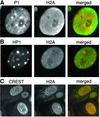
Pericentric heterochromatin becomes enriched with H2A.Z during early mammalian development
- PMID: 12660166
- PMCID: PMC152904
- DOI: 10.1093/emboj/cdg160
Pericentric heterochromatin becomes enriched with H2A.Z during early mammalian development
Abstract
Determining how chromatin is remodelled during early development, when totipotent cells begin to differentiate into specific cell types, is essential to understand how epigenetic states are established. An important mechanism by which chromatin can be remodelled is the replacement of major histones with specific histone variants. During early mammalian development H2A.Z plays an essential, but unknown, function(s). We show here that undifferentiated mouse cells of the inner cell mass lack H2A.Z, but upon differentiation H2A.Z expression is switched on. Strikingly, H2A.Z is first targeted to pericentric hetero chromatin and then to other regions of the nucleus, but is excluded from the inactive X chromosome and the nucleolus. This targeted incorporation of H2A.Z could provide a critical signal to distinguish constitutive from facultative heterochromatin. In support of this model, we demonstrate that H2A.Z can directly interact with the pericentric heterochromatin binding protein INCENP. We propose that H2A.Z functions to establish a specialized pericentric domain by assembling an architecturally distinct chromatin structure and by recruiting specific nuclear proteins.
Figures


Similar articles
-
Monoubiquitylation of H2A.Z distinguishes its association with euchromatin or facultative heterochromatin.Mol Cell Biol. 2007 Sep;27(18):6457-68. doi: 10.1128/MCB.00241-07. Epub 2007 Jul 16. Mol Cell Biol. 2007. PMID: 17636032 Free PMC article.
-
RNA interference demonstrates a novel role for H2A.Z in chromosome segregation.Nat Struct Mol Biol. 2004 Jul;11(7):650-5. doi: 10.1038/nsmb786. Epub 2004 Jun 13. Nat Struct Mol Biol. 2004. PMID: 15195148
-
H2A.Z contributes to the unique 3D structure of the centromere.Proc Natl Acad Sci U S A. 2007 Jan 9;104(2):525-30. doi: 10.1073/pnas.0607870104. Epub 2006 Dec 28. Proc Natl Acad Sci U S A. 2007. PMID: 17194760 Free PMC article.
-
Reuniting the contrasting functions of H2A.Z.Biochem Cell Biol. 2006 Aug;84(4):528-35. doi: 10.1139/o06-077. Biochem Cell Biol. 2006. PMID: 16936825 Review.
-
Quickly evolving histones, nucleosome stability and chromatin folding: all about histone H2A.Bbd.Gene. 2008 Apr 30;413(1-2):1-7. doi: 10.1016/j.gene.2008.02.003. Epub 2008 Feb 16. Gene. 2008. PMID: 18329190 Review.
Cited by
-
The H2A.Z-KDM1A complex promotes tumorigenesis by localizing in the nucleus to promote SFRP1 promoter methylation in cholangiocarcinoma cells.BMC Cancer. 2022 Nov 11;22(1):1166. doi: 10.1186/s12885-022-10279-y. BMC Cancer. 2022. PMID: 36368958 Free PMC article.
-
Histone H2A.Z inheritance during the cell cycle and its impact on promoter organization and dynamics.Nat Struct Mol Biol. 2012 Nov;19(11):1076-83. doi: 10.1038/nsmb.2424. Epub 2012 Oct 21. Nat Struct Mol Biol. 2012. PMID: 23085713
-
Analysis of mRNA abundance for histone variants, histone- and DNA-modifiers in bovine in vivo and in vitro oocytes and embryos.Sci Rep. 2019 Feb 4;9(1):1217. doi: 10.1038/s41598-018-38083-4. Sci Rep. 2019. PMID: 30718778 Free PMC article.
-
Chromatin remodelling beyond transcription: the INO80 and SWR1 complexes.Nat Rev Mol Cell Biol. 2009 Jun;10(6):373-84. doi: 10.1038/nrm2693. Epub 2009 May 8. Nat Rev Mol Cell Biol. 2009. PMID: 19424290 Free PMC article. Review.
-
Genomic organization of H2Av containing nucleosomes in Drosophila heterochromatin.PLoS One. 2011;6(6):e20511. doi: 10.1371/journal.pone.0020511. Epub 2011 Jun 27. PLoS One. 2011. PMID: 21738578 Free PMC article.
References
-
- Abbondanzo S.J., Gadi,I. and Stewart,C.L. (1993) Derivation of embryonic stem cell lines. Methods Enzymol., 225, 803–823. - PubMed
-
- Adams R.R., Wheatley,S.P., Gouldsworthy,A.M., Kandels-Lewis,S.E., Carmena,M., Smythe,C., Gerloff,D.L. and Earnshaw,W.C. (2000) INCENP binds the Aurora-related kinase AIRK2 and is required to target it to chromosomes, the central spindle and cleavage furrow. Curr. Biol., 10, 1075–1078. - PubMed
-
- Adams R.R., Carmena,M. and Earnshaw,W.C. (2001a) Chromosomal passengers and the (aurora) ABCs of mitosis. Trends Cell Biol., 11, 49–54. - PubMed
-
- Adams R.R., Eckley,D.M., Vagnarelli,P., Wheatley,S.P., Gerloff,D.L., Mackay,A.M., Svingen,P.A., Kaufmann,S.H. and Earnshaw,W.C. (2001b) Human INCENP colocalizes with the Aurora-B/AIRK2 kinase on chromosomes and is overexpressed in tumour cells. Chromosoma, 110, 65–74. - PubMed
Publication types
MeSH terms
Substances
LinkOut - more resources
Full Text Sources
Other Literature Sources
Molecular Biology Databases

