. 2003 Apr 1;22(7):1665-75.
doi: 10.1093/emboj/cdg142.

Chromosomal protein HMGN1 enhances the rate of DNA repair in chromatin

Affiliations

Chromosomal protein HMGN1 enhances the rate of DNA repair in chromatin

Yehudit Birger et al. EMBO J. .

Abstract

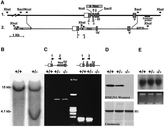
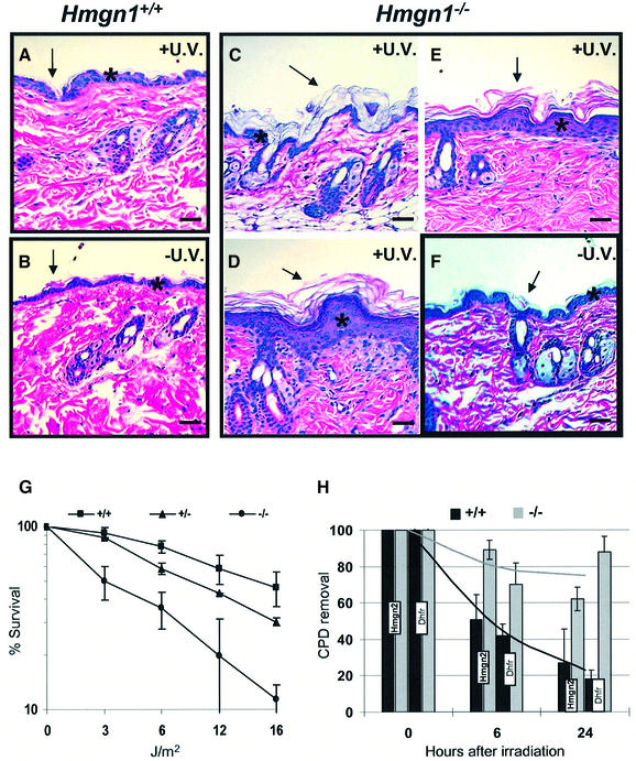
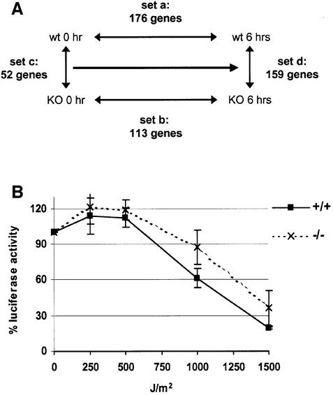
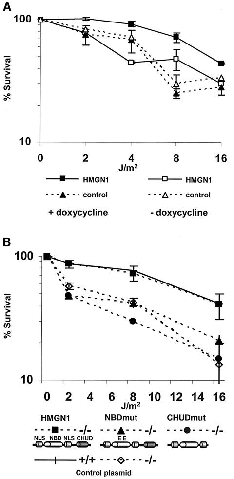
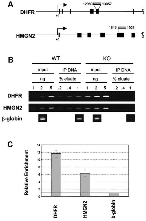
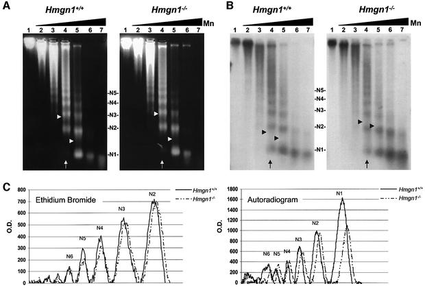
We report that HMGN1, a nucleosome binding protein that destabilizes the higher-order chromatin structure, modulates the repair rate of ultraviolet light (UV)-induced DNA lesions in chromatin. Hmgn1(-/-) mouse embryonic fibroblasts (MEFs) are hypersensitive to UV, and the removal rate of photoproducts from the chromatin of Hmgn1(-/-) MEFs is decreased as compared with the chromatin of Hmgn1(+/+) MEFs; yet, host cell reactivation assays and DNA array analysis indicate that the nucleotide excision repair (NER) pathway in the Hmgn1(-/-) MEFs remains intact. The UV hypersensitivity of Hmgn1(-/-) MEFs could be rescued by transfection with plasmids expressing wild-type HMGN1 protein, but not with plasmids expressing HMGN1 mutants that do not bind to nucleosomes or do not unfold chromatin. Transcriptionally active genes, the main target of the NER pathways in mice, contain HMGN1 protein, and loss of HMGN1 protein reduces the accessibility of transcribed genes to nucleases. By reducing the compaction of the higher-order chromatin structure, HMGN1 facilitates access to UV-damaged DNA sites and enhances the rate of DNA repair in chromatin.


Figures

None
Fig. 1. Targeted disruption of the mouse Hmgn1 gene. (A) 1, outline of the genomic sequence; 2, targeting vector and insertion sites. The targeting vector contained the TK gene with its promoter (asterisk) 5′ to Hmgn1 sequence. The Neo gene linked to HSV TK promoter (asterisk) replaced the Hmgn1 sequence from the middle of intron I to the middle of exon IV. An XbaI site that was introduced in the targeting vector and two genomic XbaI sites flanking the Hmgn1 gene were used to determine the homologous recombination, using the external probe 3′ to exon V. The primers used in PCR for genotyping the mice are indicated by the black arrowheads and numbered 1, 2 and 3. (B) Southern blot of genomic DNA from targeted ES clones digested with XbaI and hybridized with the external probe. The 15 and 4.1 kb fragments correspond to the wild-type and mutated allele, respectively. (C) Genotyping analysis by PCR. DNA samples from Hmgn1+/+, Hmgn1+/– and Hmgn –/– mice were subjected to PCR analysis with primers 1 and 2 to identify the mutated allele, and primers 1 and 3 to identify the wild-type allele. The primer positions are shown in (A). (D) Western blot analysis of 5% PCA protein extracts from Hmgn1+/+, Hmgn1+/– and Hmgn1–/– MEFs. The Coomassie Blue-stained gel below the western blot indicates that the three extracts contained equal amount of histone H1. (E) Northern blot analysis using Hmgn2 cDNA-specific probe shows equal Hmgn2 transcription in the three types of cells. Ethidium bromide staining in the lower panel shows equal amount of RNA was applied.
None
Fig. 2. Loss of HMGN1 leads to UV hypersensitivity. (A–F) Increased sensitivity of Hmgn1–/– mice to UV-B. Histology of skin after cumulative irradiation of 1200 J/m2 UV-B. (A and BHmgn1+/+ mice, (CFHmgn1–/– mice. Note the increased acanthosis (asterisk; compare E and D with F) and hyperkeratosis (arrows) in the epidermis of the Hmgn1–/– mice. (G and H) Impaired UV repair in MEFs lacking HMGN1. (G) Increased UV sensitivity of Hmgn1–/– fibroblasts. Shown is survival 72 h after irradiation with the indicated doses (see Materials and methods). (H) Decreased rate of gene-specific CPD removal in Hmgn1–/– fibroblasts. Shown is the quantitative analysis of Southern blots with probes specific for the Dhfr and Hmgn2 genes as described in Materials and methods. The 0 h point represents the initial lesion frequency. The bar graphs represent the average of three experiments.
None
Fig. 3. Intact NER in Hmgn1–/– cells. (A) Microarray analysis of gene expression in Hmgn1+/+ and Hmgn1–/– cells after UV-C irradiation at 3 J/m2 UV. The following RNA samples were compared: Array a, Hmgn1+/+ cells, before and 6 h after irradiation; Array b, Hmgn1–/– cells, before and 6 h after irradiation; Array c, non-irradiated Hmgn1+/+ and Hmgn1–/– cells; and Array d, Hmgn1+/+ and Hmgn1–/– cells 6 h after irradiation. The number of genes changed by >1.3-fold in each array experiment is indicated. The arrays contained 15 out of the 27 genes listed as NER components in the database. (B) Host-cell reactivation assay indicates that a UV-damaged luciferase reported plasmid is repaired to a similar extent in Hmgn1–/– and in Hmgn1+/+ cells.
None
Fig. 4. Rescue of UV-C sensitivity of Hmgn –/– MEFs. (A) Stable transfection with inducible HMGN1 expressing plasmids. Shown are UV survival curves of cell lines either expressing (filled squares) or not expressing (open squares) HMGN1 in the presence or absence of doxycycline. Note that addition of doxycycline reduced the UV sensitivity of cells expressing HMGN1 protein. (B) Rescue of the UV-C hypersensitivity of Hmgn1–/– cells by transient transfection of fully functional HMGN1 protein. Hmgn1–/– MEFs were transfected with vectors expressing either wild-type or mutated HMGN1 cDNA. The cells were UV irradiated 24 h after transfection and their survival rate determined 72 h later. Note that intact HMGN1, but not mutants that do not bind to nucleosomes (NBDmut) or cannot unfold chromatin (CHUDmut), rescued the UV sensitivity of the Hmgn1–/–. The schemes outline the major functional domains of Hmgn1 protein and the mutants that were transfected. NLS, nuclear localization signal; NBD, nucleosome binding domain; CHUD, chromatin unfolding domain. In the NBD mutant, two Ser residues were mutated to Glu (Prymakowska-Bosak et al., 2001). The CHUD mutant lacks the chromatin unfolding domain (Ding et al., 1997). Control, empty plasmid vector.
None
Fig. 5. Presence of HMGN1 on the chromatin of Hmgn2 and Dhfr genes. (A) Primers used to detect the genes in the ChIP assays. Black boxes indicate exons. (B) PCR analysis of chromatin immunoprecipitations from Hmgn1–/– and Hmgn1+/+ cells with antibodies to HMGN1. Note the lack of signals in the IP DNA from Hmgn1–/– cells. This analysis is not quantitative. (C) Quantification, by real time PCR analysis, of the Hmgn2 and Dhfr genes in IP from Hmgn1+/+ cells. The value for β-globin is set to 1 and the bar graphs represent the average of three experiments.
None
Fig. 6. Loss of HMGN1 decreases the accessibility of the Hmgn2 gene. Micrococcal nuclease digestions of nuclei isolated from the livers Hmgn1–/– and Hmgn1+/+ mice. (A) Ethidium bromide stain prior to Southern transfer. (B) Southern analysis with Hmgn2 specific probe. (C) Scans of lane 4 (arrow). Arrow heads indicate the length of the average oligonucleosome in the digest calculated as indicated in the methods section. The increased average oligonucleosome length in the autoradiogram of the Hmgn1–/– cells indicates slower rate of digestion of the Hmgn2 chromatin. Lanes 1–7 correspond to 0, 0.05, 0.3, 2, 11, 65 and 400 U micrococcal nuclease in the digest, respectively. N1–N5 denotes the oligonucleosomal size.

References

    1. Archer T.K., Lefebvre,P., Wolford,R.G. and Hager,G.L. (1992) Transcription factor loading on the MMTV promoter: a bimodal mechanism for promoter activation. Science, 255, 1573–1576. - PubMed
    1. Balajee A.S. and Bohr,V.A. (2000) Genomic heterogeneity of nucleotide excision repair. Gene, 250, 15–30. - PubMed
    1. Bill C.A., Grochan,B.M., Meyn,R.E., Bohr,V.A. and Tofilon,P.J. (1991) Loss of intragenomic DNA repair heterogeneity with cellular differentiation. J. Biol. Chem., 266, 21821–21826. - PubMed
    1. Bohr V.A. (1991) Gene specific DNA repair. Carcinogenesis, 12, 1983–1992. - PubMed
    1. Bohr V.A., Smith,C.A., Okumoto,D.S. and Hanawalt,P.C. (1985) DNA repair in an active gene: removal of pyrimidine dimers from the DHFR gene of CHO cells is much more efficient than in the genome overall. Cell, 40, 359–369. - PubMed