. 2003 Apr 11;327(5):1093-109.
doi: 10.1016/s0022-2836(03)00216-x.

Crystal structure of human beta-hexosaminidase B: understanding the molecular basis of Sandhoff and Tay-Sachs disease

Affiliations

Crystal structure of human beta-hexosaminidase B: understanding the molecular basis of Sandhoff and Tay-Sachs disease

Brian L Mark et al. J Mol Biol. .

Abstract

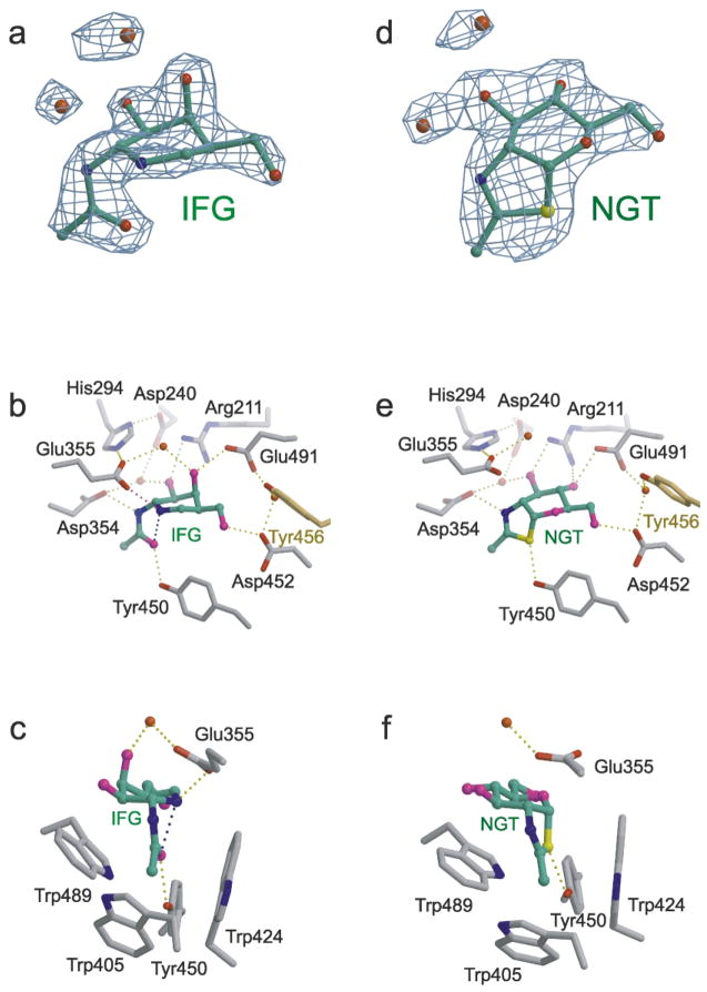
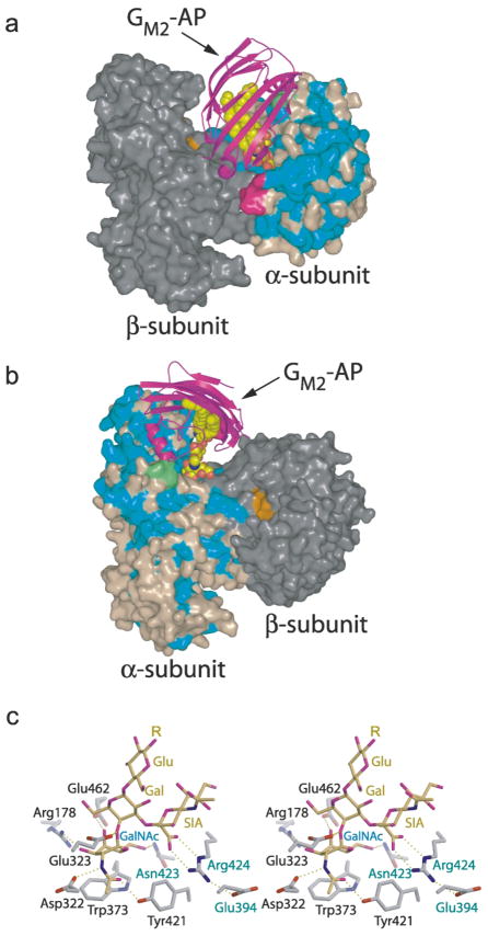
In humans, two major beta-hexosaminidase isoenzymes exist: Hex A and Hex B. Hex A is a heterodimer of subunits alpha and beta (60% identity), whereas Hex B is a homodimer of beta-subunits. Interest in human beta-hexosaminidase stems from its association with Tay-Sachs and Sandhoff disease; these are prototypical lysosomal storage disorders resulting from the abnormal accumulation of G(M2)-ganglioside (G(M2)). Hex A degrades G(M2) by removing a terminal N-acetyl-D-galactosamine (beta-GalNAc) residue, and this activity requires the G(M2)-activator, a protein which solubilizes the ganglioside for presentation to Hex A. We present here the crystal structure of human Hex B, alone (2.4A) and in complex with the mechanistic inhibitors GalNAc-isofagomine (2.2A) or NAG-thiazoline (2.5A). From these, and the known X-ray structure of the G(M2)-activator, we have modeled Hex A in complex with the activator and ganglioside. Together, our crystallographic and modeling data demonstrate how alpha and beta-subunits dimerize to form either Hex A or Hex B, how these isoenzymes hydrolyze diverse substrates, and how many documented point mutations cause Sandhoff disease (beta-subunit mutations) and Tay-Sachs disease (alpha-subunit mutations).


Figures

Figure 1
Figure 1
Proposed catalytic mechanism for family 20 β-hexosaminidase. The Hex B residues Glu355 (Hex A; Glu323) and Asp354 (Hex A; Asp322) are shown. Glu355 acts as a general acid–base residue, whereas Asp354 acts primarily to help in orienting the C2-acetamido group into a position for nucleophilic attack and subsequently to stabilize the positive charge on the oxazolinium ion intermediate. No attempt has been made to indicate the true positions of these residues. The hydroxyl groups and C6 have been removed from the pyranose ring of the substrate for clarity.
Figure 2
Figure 2
Ribbon diagram of human β-hexosaminidase B. The β-subunits of the Hex B homodimer are colored with domain I in green and domain II in blue (the eight parallel strands of the β-barrel of domain II is colored sky blue). What appear to be common structural features of family 20 glycosidases is the absence of regular α-helices at positions α5 and α7 of the (β/α)8-barrel structure of domain II and an additional C-terminal helix following helix α8. This additional helix packs between domains I and II, spatially orienting the two domains relative to each other. Helix α7 consists of only two turns and is part of an extended loop that forms a major portion of the dimer interface. The subunits are related at the dimer interface by a crystallographic 2-fold symmetry axis running perpendicular to the page. The N and C termini created as a result of post-translational processing are numbered by residue. The labels N and C denote the extreme N (residue 55) and C (residue 552) termini visible within the electron density. The disulfide bonds Cys91-Cys137, Cys309-Cys360 and Cys534-Cys551 are drawn in brown, magenta and yellow, respectively. The analogue of the reaction intermediate NAG-thiazoline, bound in the active site of each subunit is drawn as a space-filling model with carbon atoms in gray, oxygen in red, nitrogen in blue and sulfur in yellow. The active sites of each subunit are located 37 Å apart. All ribbon diagrams were drawn with Molscript and rendered with Raster3D unless otherwise indicated.
Figure 3
Figure 3
Electrostatic potential surface map and dimer interface of human Hex B. (a) A solvent-accessible surface, drawn over one β-subunit and colored with regions of positive charge in blue and negative charge in red, reveals an overall negative charge about the active site. Note, however, that the electrostatic surface was calculated using the following charge profile only: Lys atom Nζ (charge 1.0); Arg atom NH1 (charge 0.5) and NH2 (charge 0.5); Glu atom Oε1 (charge −0.5) and Oε2 (charge −0.5); Asp atom Oδ1 (charge −0.5) and Oδ1 (charge −0.5); His atom Nδ1 (charge 0.5) and Nε2 (charge 0.5); OXT (charge −1.0). Due to the acidic environment of the lysosome, the electrostatic surface potential of Hex B in the lysosome may be slightly different from what is represented here, potentially being less negatively charged about the active site due to protonation of Glu and Asp carboxyl groups (surface created using the program GRASP). The other subunit of the homodimer is represented by a ribbon diagram with domain I in green and the catalytic (β/α)8 domain II in yellow. The intermediate analogue NAG-thiazoline, bound in the active site of each subunit is shown as a space-filling model with carbon atoms in gray, oxygen in magenta, nitrogen in blue and sulfur in yellow. (b) Surface rendering of a single β-subunit showing the extensive surface area buried at the dimer interface as determined using the CNS program. Polar side-chains are colored blue, hydrophobic side-chains in yellow, backbone atoms in forest green, charged residues in magenta and residues not involved in dimerization are colored gray. The active site pocket is colored red ((b) was drawn using the program PyMOL). (c) Active site residues (gray) stabilized by interactions from residues of the partnering subunit (yellow). The 2-fold symmetry at the dimer interface results in both active sites experiencing the same stabilizing effects from the associated monomer. The crystallographically determined position of GalNAc–isofagomine (IFG) in the active site of each subunit demonstrates that four of the six hydrogen bonds between the enzyme and inhibitor depend on stabilizing interactions from the partnering subunit. In the absence of the protein–protein interactions that are formed upon dimerization, Arg211, Glu491, Asp452 and Tyr450 are most likely too unstructured to be catalytically acitve.
Figure 4
Figure 4
Pair-wise sequence alignment and secondary structure of subunits α and β. Residues colored in light blue are removed during post-translational processing, and residues in italics compose the ER signal peptides of each subunit (Table 1). Sites (N-X-S/T) known to contain N-linked oligosaccharides are underlined, and glycan sites that receive the mannose-6-phosphate lysosomal targeting moiety are doubly underlined (Table 1). Primary sequence corresponding to the mature, lysosomal αp and βp chains are surrounded by square brackets, sequence comprising chains αm and βb are in curly brackets, and the sequence for chain βa is surrounded by normal brackets. Secondary structural elements are as follows: α-helices are drawn as green boxes, β-strands are drawn as blue arrows and disulfide bridges are shown by blue–gray lines connecting Cys residues. Residues boxed in yellow are involved in subunit dimerization as determined from the Hex B crystal structure and also predicted for the Hex A isozyme. The unique mature α-subunit loops 280–283 (GSEP) and 396–398 (IPV) are colored magenta and are predicted to interact directly with the bound activator protein. β-Subunit point mutations known to cause GM2-gangliosidosis are indicated directly above the β-subunit sequence in purple.
Figure 5
Figure 5
Hex B in complex with the transition state mimic GalNAc-isofagomine (IFG) (a–c) or the intermediate analogue NAG-thiazo-line (NGT) (d–f). (a) Unrefined 2.2 Å resolution sigma-A weighted |Fo| − |Fc|, αc electron density map containing the refined model of IFG bound in the active site of a Hex B β-subunit (αc are the calculated phases and |Fo| and |Fc| are the measured and calculated structure factor amplitudes, respectively). Carbon atoms of IFG are colored green, nitrogen blue, oxygen red. (b) The β-subunit active site showing the extensive hydrogen-bonding interactions between IFG and the enzyme. The magenta hydrogen bond between Glu355 and the ring nitrogen atom of IFG is believed to compensate for the missing hydrogen bond that occurs between an isofagomine inhibitor and a “normal” β-retaining glycosidase containing an enzyme nucleophile., Tyr456 (yellow) comes into the active site from the partnering subunit to stabilize the position of a water molecule linking Asp452 and Glu491, two residues involved in determining substrate specificity. (c) Tryptophan residues create a hydrophobic pocket in the active site into which the C2-acetamido group becomes appropriately positioned for intramolecular nucleophilic attack at the anomeric center of the terminal sugar molecule being removed from the substrate. The water molecule located above the β-face of the azasugar ring of IFG may represent the incoming water that undergoes base catalyzed (activated by Glu355) nucleophilic attack at the anomeric center of the cyclized intermediate to produce a product with retained configuration. (d) Unrefined 2.5 Å resolution sigma-A weighted |Fo| − |Fc|, αc electron density map containing the refined model of NGT bound in the active site of a Hex B β-subunit (αc are the calculated phases and |Fo| and |Fc| are the measured and calculated structure factor amplitudes, respectively). Carbon atoms of NGT are colored green, nitrogen blue, oxygen red and sulfur yellow. (e) Hydrogen-bonding interactions within the active site that stabilize the cyclized enzyme intermediate. Tyr456 (yellow) comes into the active site from the partnering subunit. (f) Once cyclization has occurred, the enzyme intermediate is protected from solvolysis via unwanted pathways within the tryptophan-lined pocket. A water molecule, activated by Glu355, can only attack from above the β-face, ensuring net retention of anomeric configuration of the product.
Figure 6
Figure 6
Predicted model of human Hex A–GM2–activator quaternary complex. (a and b) Two views of the predicted quaternary complex. Residues of the α-subunit identical to those of the β-subunit are colored blue, non-identical residues are colored light brown. Most of the conserved amino acids in the α and β-subunits are located in (β/α)8-barrel of domain II. The β-subunit is colored gray, with residues of the active site distinguished in orange. The GM2–activator protein complex (GM2–AP) docks into a large groove between the two subunits so that the terminal non-reducing GalNAc sugar on GM2 can be presented to the α-subunit active site and removed. Two surface loops (magenta and green), present only on the α-subunit, interact with the docked activator protein and appear to be involved in creating a docking site unique to the α-subunit. The magenta colored loop is proteolytically removed from the β-subunit during post-translational processing and may represent a modification that regulates the metabolic function of this subunit. (c) Model of the GM2 oligosaccharide (yellow) bound to the α-subunit active site (gray). The distorted boat conformation of the terminal GalNAc to be removed (Gal, labeled in blue) and the pseudoaxial orientation of the scissile bond and leaving group are based on crystallographic observations of the Michaelis complex of chitobiose bound to SmCHB. By incorporating these conformational restraints into the model, only one reasonable position could be found for the sialic acid residue (labeled SIA) within the active site pocket. Once positioned, the negatively charged carboxylate of the sialic acid, which can only be accommodated by the α-subunit, was found to come within hydrogen bonding distance of Arg424, a positively charged residue that is unique to the α-subunit (the β-subunit contains a Leu at this position). αGlu394 and αAsn423 (which are both Asp residues in the β-subunit) are believed to help hold Arg424 into position. Arg424, in turn, stabilizes the negatively charged caboxylate of the sialic acid of the substrate via electrostatic and hydrogen-bonding interactions. The general acid–base residue, Glu323 (Glu355 in the β-subunit), can be seen interacting with the glycosidic oxygen atom of the scissile bond.

References

    1. Watanabe K. Biochemical studies on carbohydrates. J Biochem (Tokyo) 1936;24:297.
    1. Tay W. Symmetrical changes in the region of the yellow spot in each eye of an infant. Trans Ophthalmol Soc UK. 1881;1:155–157.
    1. Gravel RA, Clarke JTR, Kaback MM, Mahuran D, Sandhoff K, Suzuki K. The GM2 gangliosidoses. In: Scriver CR, Beaudet AL, Sly WS, editors. The Metabolic and Molecular Bases of Inherited Disease. 7. Vol. 2. Vol. 3 McGraw-Hill; New York: 1995. pp. 2839–2879.
    1. Mahuran DJ. Biochemical consequences of mutations causing the GM2 gangliosidoses. Biochim Biophys Acta. 1999;1455:105–138. - PubMed
    1. Mahuran DJ. The GM2 activator protein, its roles as a co-factor in GM2 hydrolysis and as a general glycolipid transport protein. Biochim Biophys Acta. 1998;1393:1–18. - PubMed

Publication types

Grants and funding