. 2003 Apr;77(8):4502-15.
doi: 10.1128/jvi.77.8.4502-4515.2003.

CXCL10 production from cytomegalovirus-stimulated microglia is regulated by both human and viral interleukin-10

Affiliations

CXCL10 production from cytomegalovirus-stimulated microglia is regulated by both human and viral interleukin-10

Maxim C-J Cheeran et al. J Virol. 2003 Apr.

Abstract

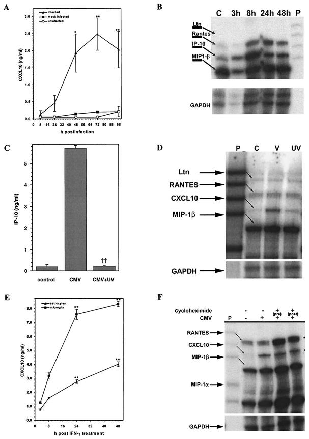
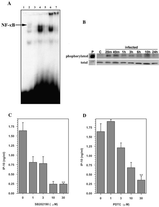
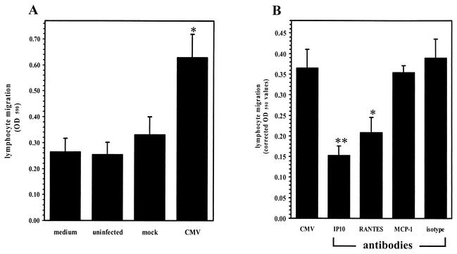
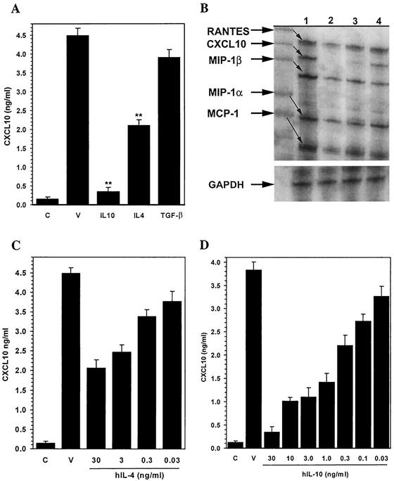
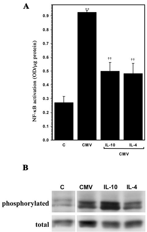
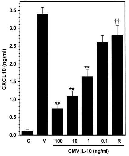
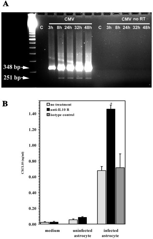
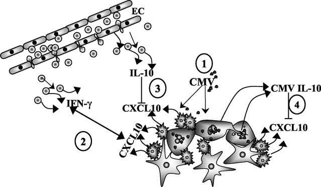
Glial cells orchestrate immunocyte recruitment to focal areas of viral infection within the brain and synchronize immune cell functions through a regulated network of cytokines and chemokines. Since recruitment of T lymphocytes plays a critical role in resolving cytomegalovirus (CMV) infection, we investigated the production of a T-cell chemoattractant, CXCL10 (gamma interferon-inducible protein 10) in response to viral infection of human glial cells. Infection with CMV was found to elicit the production of CXCL10 from primary microglial cells but not from astrocytes. This CXCL10 expression was not dependent on secondary protein synthesis but did require the phosphorylation of p38 mitogen-activated protein (MAP) kinase. In addition, migration of activated lymphocytes toward supernatants from CMV-stimulated microglial cells was partially suppressed by anti-CXCL10 antibodies. Since regulation of central nervous system inflammation is essential to allow viral clearance without immunopathology, microglial cells were then treated with anti-inflammatory cytokines. CMV-induced CXCL10 production from microglial cells was suppressed following treatment with interleukin-10 (IL-10) and IL-4 but not following treatment with transforming growth factor beta. The IL-10-mediated inhibition of CXCL10 production was associated with decreased CMV-induced NF-kappa B activation but not decreased p38 MAP kinase phosphorylation. Finally, CMV infection of fully permissive astrocytes resulted in mRNA expression for the viral homologue to human IL-10 (i.e., cmvIL-10 [UL111a]) in its spliced form and conditioned medium from CMV-infected astrocytes inhibited virus-induced CXCL10 production from microglial cells through the IL-10 receptor. These findings present yet another mechanism through which CMV may subvert host immune responses.


Figures

FIG.1.
FIG.1.
Microglial cells produce CXCL10 in response to stimulation with CMV. (A) Induction of CXCL10 protein. Microglial cells (2 × 10⁵) were exposed to CMV AD169 at an MOI of 5 TCID50 (infected) or to virus-free HFF cell extracts (mock infected) or medium alone (uninfected) for 8, 24, 48, 72, and 96 h prior to measurement of supernatant CXCL10 levels by ELISA. Data are expressed as mean (± SEM) CXCL10 levels from pooled data obtained during three separate experiments with microglial cells isolated from three different brain specimens. *, P < 0.05; **, P < 0.01 (versus mock-infected control cells). (B) Kinetics of chemokine mRNA induction in response to CMV. Total RNA was extracted from CMV-exposed microglial cells at 3, 8, 24, and 48 h, and 4 μg was used in the multiprobe chemokine RPA in accordance with the manufacturer's (Pharmingen) instructions. Lane C, uninfected microglial cell RNA; lane P, probe alone. Ltn, lymphotactin. GAPDH, glyceraldehyde-3-phosphate dehydrogenase. (C) UV inactivation decreases CXCL10 induction in microglial cells. Microglial cells (2 × 10⁵) were stimulated with UV-inactivated CMV AD169 (equivalent to an MOI of 5 TCID50). CXCL10 levels were measured by ELISA at 72 h p.i.. Data are presented as the mean ± the SEM of pooled samples from three separate experiments. ††, P < 0.01 versus infected cells. (D) Decreased CXCL10 mRNA expression in microglial cells by UV-inactivated CMV. Total RNA from microglial cells stimulated with replication-competent (V) or UV-inactivated (UV) CMV or from uninfected cells (C) was analyzed by RPA at 24 h p.i. (E) IFN-γ (200 U/ml)-exposed microglial cells (microglia) were used as a positive control. CXCL10 production was also examined following IFN-γ treatment of astrocytes (2 × 10⁵ astrocytes). The data shown are the mean ± the SEM of pooled data from three separate experiments with glial cells obtained from two different brain specimens. **, P < 0.01 versus untreated cells. (F) Microglial cells were treated with cycloheximide (10 μg/ml) 30 min before (pre) or 6 h after (post) the addition of CMV. Total RNA was extracted from cycloheximide-treated and control cultures at 18 h p.i. and analyzed for CXCL10 mRNA by RPA.
FIG. 2.
FIG. 2.
Inhibitors of signal transduction block CMV-induced CXCL10 production. (A) Mobility shift assay with nuclear extracts from microglial cells stimulated with CMV (MOI of 5 TCID50). Nuclear extracts (2 μg) were probed for NF-κB binding activity. Lanes: 1, probe alone; 2, HeLa cell extract (positive control for NF-κB binding); 3, uninfected microglial cell extract; 4, CMV-stimulated microglial cell extract, 2 h p.i.; 5, CMV-stimulated microglial cell extract, 24 h p.i.; 6, same extract as lane 4 supershifted with anti-p65 antibody; 7, same extract as lane 5 supershifted with anti-p65 antibody. (B) Kinetics of phosphorylation of microglial cell p38 MAP kinase in response to CMV. At the indicated times p.i., whole-cell lysates were obtained for analysis of phosphorylated p38 MAP kinase and total p38 by Western blot assay. Lane P, positive control for phosphorylated p38; lane C, extract obtained from uninfected microglial cells. (C and D) Microglial cells were treated with SB202190 (a p38 MAP kinase inhibitor) (C) or PDTC (an inhibitor of transcription factors) (D) at the indicated concentrations (1 to 30 μM) for 30 min and 2 h prior to addition of CMV, respectively. Culture supernatants were harvested 72 h p.i., and CXCL10 levels were quantified by ELISA. Data are presented as the mean ± the SEM of triplicate samples and are representative of three or more experiments performed with cells derived from different brain specimens. **, P < 0.01 versus the untreated control.
FIG. 3.
FIG. 3.
Activated lymphocytes migrate toward supernatants from CMV-infected microglial cells. (A) Cell migration was assessed in a 96-well chemotaxis chamber (Neuro Probe). Supernatants from CMV-stimulated microglial cells (48 h p.i.) were added to the lower wells, and activated lymphocytes (2 × 10⁶ cells/well) in RPMI were added to the upper wells of the chemotaxis chamber. After 3 h of incubation at 37°C, the cells that had migrated to the lower plate were collected by centrifugation and quantified by spectrophotometry with an MTT assay. Medium, medium alone; uninfected, supernatants from uninfected microglial cells; mock, supernatants from mock-infected microglial cells; CMV, supernatants from CMV-infected microglial cells. Data are presented as the mean (± the SEM) of pooled data from three separate experiments utilizing glial cells from different donor specimens. *, P < 0.05 versus mock-infected supernatants. OD 590, optical density at 590 nm. (B) Inhibition of T-cell migration by anti-CXCL10 antibodies. Supernatants collected from CMV-infected microglial cells were treated (30 min) with 10 μg of specific antichemokine antibody per ml prior to assessment of chemotaxis. IP10, anti-CXCL10 antibody; RANTES, anti-RANTES antibody; isotype, isotype-matched control antibody. Data are presented as the mean (± the SEM) corrected optical density at 590 nm (migration toward sample supernatants minus random migration toward medium) from pooled data of three separate experiments with glial cells obtained from three different brain cell specimens. *, P < 0.05; **, P < 0.01 (versus untreated CMV-stimulated supernatant).
FIG. 4.
FIG. 4.
Select anti-inflammatory cytokines regulate CMV-induced CXCL10 production by human microglial cells. (A) Anti-inflammatory cytokine-mediated inhibition of CXCL10 production as measured in microglial cell supernatants by ELISA. C, uninfected microglial cell control; V, supernatants obtained from CMV-infected microglial cells; IL-10, supernatants from cells treated with IL-10 (30 ng/ml overnight) prior to CMV infection; IL-4, IL-4 treatment prior to infection; TGF-β, supernatants from cells treated with TGF-β (30 ng/ml overnight) prior to CMV infection. **, P < 0.01 versus untreated, CMV-infected microglial cells. (B) IL-10- and IL-4-mediated inhibition of CXCL10 mRNA. A chemokine RPA was performed. Lanes: 1, RNA extracted from CMV-infected microglial cells; 2, uninfected microglial cell RNA; 3, RNA extracted from microglial cells treated with IL-10 (30 ng/ml overnight) prior to CMV infection; 4, RNA extracted from microglial cells treated with IL-4 (30 ng/ml overnight) prior to CMV infection. GAPDH, glyceraldehyde-3-phosphate dehydrogenase. (C and D) Concentration-response effects of IL-4 (C) and IL-10 (D) on CMV-induced CXCL10 production. Microglial cells were treated overnight with the indicated concentrations of IL-4 or IL-10 (0.03 to 30 ng/ml) prior to CMV infection. C, uninfected microglial cell control; V, CMV-infected microglial cells. Data are expressed as mean (± SEM) CXCL10 levels from pooled data obtained from three separate experiments with microglial cells isolated from three different brain cell specimens.
FIG. 5.
FIG. 5.
IL-10 treatment of microglial cells is associated with decreased CMV-induced activation of NF-κB but not phosphorylation of p38 MAP kinase. (A) Primary human microglial cells were treated overnight with IL-10 (30 ng/ml) or IL-4 (30 ng/ml) and then stimulated with sucrose-purified CMV. Activation of microglial cell NF-κB was quantified 1.5 h p.i. with an NF-κB ELISA. **, P < 0.01 versus unstimulated microglial cells; ††, P < 0.01 versus stimulated microglial cells. OD, optical density. (B) Microglial cells maintained in serum-free medium were treated with the indicated anti-inflammatory cytokine prior to viral (CMV) infection or addition of medium alone (uninfected). Whole-cell lysates were obtained for analysis of phosphorylated p38 MAP kinase (phosphorylated) and total p38 (total) by Western blot assay. C, extract obtained from uninfected microglial cells.
FIG. 6.
FIG. 6.
Microglial cell-mediated CXCL10 production is downregulated by CMV IL-10. Concentration-response effect of CMV IL-10 on virus-induced CXCL10 production by microglial cells. Microglial cells were treated overnight with the indicated concentration of recombinant CMV IL-10 (0.1 to 100 ng/ml) prior to infection with CMV AD169. ELISA was used to quantify CXCL10 levels in the infected-cell supernatants. C, uninfected microglial cell control; V, CMV-infected microglial cells; R, microglial cells treated with an antibody to the human IL-10 receptor (15 μg/ml) prior to CMV IL-10 (100 ng/ml) treatment. Data are expressed as mean (± SEM) CXCL10 levels from pooled data obtained from three separate experiments with microglial cells isolated from three different brain specimens. **, P < 0.01 versus untreated CMV-infected microglial cells; ††, P < 0.01 versus microglial cells treated with 100 ng of CMV IL-10 per ml.
FIG. 7.
FIG. 7.
cmvIL-10 is expressed during viral replication in human astrocytes. (A) mRNA expression. PCR analysis was performed on RNA extracted from uninfected astrocytes (lane C) and astrocytes at 3, 8, 24, 32, and 48 h following infection with CMV AD169 (MOI of 1 TCID50). Identical samples were also subjected to PCR amplification without RT treatment (no RT). The calculated size of the genomic-length PCR product is 348 bp, and that of the product corresponding to the spliced mRNA is 251 bp. cmvIL-10 mRNA was not present at 3 h p.i. but was detected by 8 h p.i. The gel shown is representative of three independent experiments with astrocytes from different brain specimens. (B) Conditioned medium from CMV-infected astrocytes inhibits microglial cell CXCL10 production through the IL-10 receptor. Microglial cells were treated with anti-IL-10 receptor antibody or isotype control antibody (15 μg/ml, 30 min) prior to the addition of medium alone or medium from uninfected or CMV-infected astrocytes (MOI of 5 TCID50). CXCL10 production was quantified by ELISA 72 h post supernatant addition. Data, expressed as the mean ± the SEM, are representative of four independent experiments with glial cells from different brain specimens. *, P < 0.05 versus untreated astrocyte supernatants.
FIG. 8.
FIG. 8.
Model for CMV subversion of neuroimmune responses through inhibition of CXCL10 production. (Step 1) CMV productively infects astrocytes and stimulates microglial cells to produce CXCL10. (Step 2) Activated T lymphocytes respond to this microglial cell-produced CXCL10 and migrate toward foci of infection in the CNS. Through the production of soluble factors such as IFN-γ, these lymphocytes work to control viral replication and spreading. (Step 3) Additional lymphocytes make available anti-inflammatory cytokines such as IL-10 that suppress CXCL10 production by microglial cells and prevent excessive inflammatory brain damage. (Step 4) CMV exploits these immunosuppressive effects by expressing a viral homologue of IL-10 (i.e., CMV IL-10) that functions to dampen CXCL10 production and possibly reduce lymphocyte-mediated viral clearance.

References

    1. Aloisi, F. 2001. Immune function of microglia. Glia 36:165-179. - PubMed
    1. Arribas, J. R., G. A. Storch, D. B. Clifford, and A. C. Tselis. 1996. Cytomegalovirus encephalitis. Ann. Intern. Med. 125:577-587. - PubMed
    1. Asensio, V. C., and I. L. Campbell. 1997. Chemokine gene expression in the brains of mice with lymphocytic choriomeningitis. J. Virol. 71:7832-7840. - PMC - PubMed
    1. Asensio, V. C., J. Maier, R. Milner, K. Boztug, C. Kincaid, M. Moulard, C. Phillipson, K. Lindsley, T. Krucker, H. S. Fox, and I. L. Campbell. 2001. Interferon-independent, human immunodeficiency virus type 1 gp120-mediated induction of CXCL10/IP-10 gene expression by astrocytes in vivo and in vitro. J. Virol. 75:7067-7077. - PMC - PubMed
    1. Chang, L., and M. Karin. 2001. Mammalian MAP kinase signalling cascades. Nature 410:37-40. - PubMed

Publication types

MeSH terms

LinkOut - more resources