. 2003 Apr;77(8):4646-57.
doi: 10.1128/jvi.77.8.4646-4657.2003.

Ability of the matrix protein of vesicular stomatitis virus to suppress beta interferon gene expression is genetically correlated with the inhibition of host RNA and protein synthesis

Affiliations

Ability of the matrix protein of vesicular stomatitis virus to suppress beta interferon gene expression is genetically correlated with the inhibition of host RNA and protein synthesis

Maryam Ahmed et al. J Virol. 2003 Apr.

Abstract

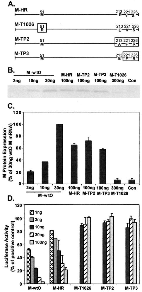
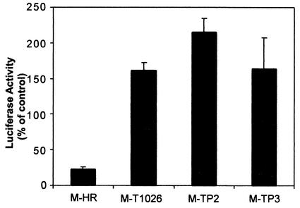
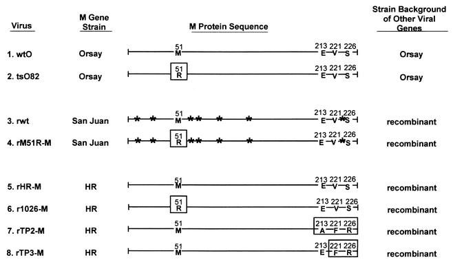
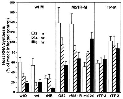
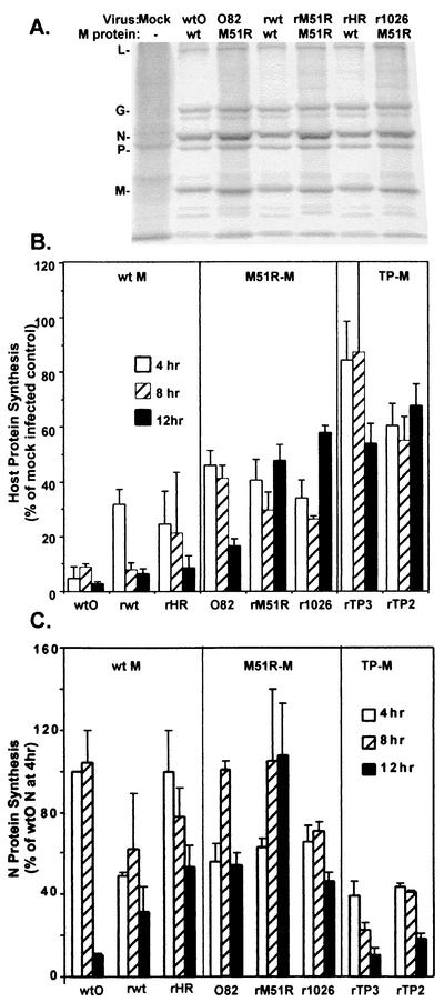
The vesicular stomatitis virus (VSV) matrix (M) protein plays a major role in the virus-induced inhibition of host gene expression. It has been proposed that the inhibition of host gene expression by M protein is responsible for suppressing activation of host interferon gene expression. Most wild-type (wt) strains of VSV induce little if any interferon gene expression. Interferon-inducing mutants of VSV have been isolated previously, many of which contain mutations in their M proteins. However, it was not known whether these M protein mutations were responsible for the interferon-inducing phenotype of these viruses. Alternatively, mutations in other genes besides the M gene may enhance the ability of VSV to induce interferons. These hypotheses were tested by transfecting cells with mRNA expressing wt and mutant M proteins in the absence of other viral components and determining their ability to inhibit interferon gene expression. The M protein mutations were the M51R mutation originally found in the tsO82 and T1026R1 mutant viruses, the double substitution V221F and S226R found in the TP3 mutant virus, and the triple substitution E213A, V221F, and S226R found in the TP2 mutant virus. wt M proteins suppressed expression of luciferase from the simian virus 40 promoter and from the beta interferon (IFN-beta) promoter, while M proteins of interferon-inducing viruses were unable to inhibit luciferase expression from either promoter. The M genes of the interferon-inducing mutants of VSV were incorporated into the wt background of a recombinant VSV infectious cDNA clone. The resulting recombinant viruses were tested for their ability to activate interferon gene expression and for their ability to inhibit host RNA and protein synthesis. Each of the recombinant viruses containing M protein mutations induced expression of a luciferase reporter gene driven by the IFN-beta promoter and induced production of interferon bioactivity more effectively than viruses containing wt M proteins. Furthermore, the M protein mutant viruses were defective in their ability to inhibit both host RNA synthesis and host protein synthesis. These data support the idea that wt M protein suppresses interferon gene expression through the general inhibition of host RNA and protein synthesis.


Figures

FIG. 1.
FIG. 1.
Effect of wt and mutant M proteins on expression of luciferase from the SV40 promoter. (A) Diagram representing sequences of M proteins from IFN-inducing mutant viruses of the HR strain of VSV. M-HR is the wt M protein of the HR strain. Mutations in the M-T1026, M-TP2, and M-TP3 proteins are indicated by boxes. (B) Representative image of M proteins expressed from cells transfected with wt and mutant M mRNAs. L cells were transfected with the indicated amounts of wt and mutant M mRNAs for 5 h. Cells transfected with pGL3 plasmid DNA alone were used as a negative control. Cells were labeled with [35S]methionine (200 μCi/ml) for 1 h, and cell extracts were prepared. Extracts were immunoprecipitated with the anti-M protein monoclonal antibody 23H12 and processed for SDS-PAGE and phosphorimaging. (C) Quantitation of labeled M proteins. Results are expressed as the percentage of M protein expressed in cells transfected with 30 ng of wtO mRNA. Data are the means ± standard errors of the means for four experiments. (D) Effect of wt and mutant M proteins on expression of luciferase from the SV40 promoter. HeLa cells were transfected with the indicated amounts of in vitro-transcribed M mRNA together with 250 ng of pGL3 plasmid DNA containing a luciferase gene driven by the SV40 promoter. At 24 h posttransfection, cells were harvested and luciferase activity was measured. Data are presented as the percentage of the activity of controls transfected with pGL3 plasmid DNA in the absence of M mRNA and are the means ± standard errors of the means for eight independent experiments.
FIG. 2.
FIG. 2.
Effect of wt and mutant M proteins on expression of luciferase from the IFN-β promoter. HeLa cells were transfected with 1 μg of pβlux encoding luciferase expressed from the IFN-β promoter, together with 100 ng of wt, TP2, and TP3 M mRNA or 300 ng of T1026 M mRNA. At 16 h posttransfection, cell extracts were prepared, and luciferase activity was measured. Luciferase activities are expressed as a percentage of the activity in cells transfected with pβlux alone and are the means ± standard errors of the means for four independent experiments.
FIG. 3.
FIG. 3.
Viruses used in this study. The diagram represents the sequences of the M proteins of the viruses used in this study. Mutations in the M proteins are indicated by boxes. The tsO82 virus (number 2) is a naturally occurring mutant of the Orsay strain of VSV (number 1) containing the M51R mutation (11). The remaining viruses are recombinants isolated from VSV infectious cDNA clones, which differ only in their M genes. The M genes of the original recombinant wt (rwt) virus (3) and rM51R-M mutant (4) are derived from the San Juan strain (27, 42). The viruses containing the wt M protein from the HR strain (rHR-M virus) (5), the M51R mutation in the HR M protein (r1026-M virus) (6), and the TP2 and TP3 mutations in the HR M protein (7 and 8) were generated for this study. Sites of amino acid differences between the San Juan and HR strains are indicated by asterisks.
FIG. 4.
FIG. 4.
Effect of viruses containing wt or mutant M proteins on the activity of the IFN-β promoter. HeLa cells were transfected with 1 μg of pβlux plasmid DNA encoding luciferase under control of the IFN-β promoter. At 24 h posttransfection, cells were infected with viruses containing wt or mutant M proteins at a multiplicity of 20 PFU/cell. Cells were harvested at 3, 6, and 9 h postinfection, and luciferase activity was determined. Cells were transfected with pβlux DNA and mock infected as negative controls (Con), and cells were transfected with pβlux DNA and then treated with poly(I)-poly(C) (pI:pC) as positive controls. Data are expressed as a percentage of the luciferase activity expressed by uninfected cells transfected with 250 ng of pGL3 plasmid DNA to detect constitutive luciferase activity from the SV40 promoter. Data shown are means ± standard errors of the means for four independent experiments.
FIG. 5.
FIG. 5.
IFN bioactivity produced by cells infected with wt and mutant M protein viruses. HeLa cells were incubated overnight at 37°C with serial dilutions of supernatants (100 μl) collected from HeLa (A) and PC-3 (B) cells infected with wt and mutant viruses. The samples were aspirated, and cells were challenged with wt VSV at 2.24 × 10⁴ PFU/ml in 100 μl of medium. Cells were incubated overnight at 37°C, medium was aspirated, and cells were fixed and stained with crystal violet. Absorbance was read at 550 nm on an ELISA reader. The IFN concentration (in international units per milliliter) was quantitated by comparing results to those in cells incubated with serial fivefold dilutions of an IFN standard. Data shown are means ± standard errors of the means for three independent experiments.
FIG. 6.
FIG. 6.
Inhibition of host RNA synthesis by viruses containing wt or mutant M proteins. HeLa cells were infected with viruses containing wt or mutant M proteins at a multiplicity of 20 PFU/cell. At 2, 4, and 6 h postinfection, cells were labeled with [3H]uridine (20 μCi/ml) for 30 min. Cells were lysed in SDS-lysis buffer, and aliquots were precipitated with trichloroacetic acid to measure acid-insoluble radioactivity. Parallel samples were incubated in the presence of actinomycin D, so that only viral RNA would be labeled. The rate of host RNA synthesis was calculated by subtracting the radioactivity in viral RNA from the total radioactivity. Data are expressed as a percentage of the uninfected cell control and are means ± standard errors of the means for five experiments.
FIG. 7.
FIG. 7.
Inhibition of host protein synthesis by viruses containing wt or mutant M proteins. HeLa cells were infected with viruses containing wt or mutant M proteins at a multiplicity of 20 PFU/cell or were mock infected as a control. Cells were labeled with a 15-min pulse of [35S]methionine (100 μCi/ml) at 4, 8, and 12 h postinfection. Lysates were subjected to SDS-PAGE, and labeled proteins were quantitated by phosphorimaging. (A) Representative image from analysis of viruses containing wt or M51R mutant M proteins at 8 h postinfection. Positions of viral proteins are indicated on the left. (B) Host protein synthesis was determined from images similar to that in panel A in regions of the gel devoid of viral proteins between the L and G proteins and between the P and M proteins. Results are shown as a percentage of the mock-infected control and are the mean ± standard error of the mean of four independent experiments. (C) Effect of M protein mutations on viral protein synthesis. HeLa cells infected with viruses containing wt or mutant M proteins were labeled with [35S]methionine, and the labeled proteins were analyzed by SDS-PAGE and phosphorimaging as described in the legend for Fig. 4. The labeled M proteins in images similar to those shown in Fig. 4A were quantitated and are expressed as a percentage of the wtO M protein labeled at 4 h postinfection. Data are the mean ± standard error of the mean of four experiments.
FIG. 8.
FIG. 8.
Single-cycle growth analysis. HeLa cells were infected with viruses containing wt or mutant M proteins at a multiplicity of 20 PFU/cell. At 1 h postinfection, the medium was removed, and cells were washed twice. Fresh medium was added to the infected cells, and a small aliquot of the supernatant was removed at the indicated times postinfection to determine the amount of progeny virus by plaque assay. Data are the average of two independent experiments.

References

    1. Ahmed, M., and D. S. Lyles. 1998. Effect of vesicular stomatitis virus matrix protein on transcription directed by host RNA polymerases I, II, and III. J. Virol. 72:8413-8419. - PMC - PubMed
    1. Ahmed, M., and D. S. Lyles. 1997. Identification of a consensus mutation in M protein of vesicular stomatitis virus from persistently infected cells that affects inhibition of host-directed gene expression. Virology 237:378-383. - PubMed
    1. Alcami, A., J. A. Symons, and G. L. Smith. 2000. The vaccinia virus soluble alpha/beta interferon (IFN) receptor binds to the cell surface and protects cells from the antiviral effects of IFN. J. Virol. 74:11230-11239. - PMC - PubMed
    1. Black, B. L., G. Brewer, and D. S. Lyles. 1994. Effect of vesicular stomatitis virus matrix protein on host-directed translation in vivo. J. Virol. 68:555-560. - PMC - PubMed
    1. Black, B. L., and D. S. Lyles. 1992. Vesicular stomatitis virus matrix protein inhibits host cell-directed transcription of target genes in vivo. J. Virol. 66:4058-4064. - PMC - PubMed

Publication types

LinkOut - more resources