. 2003 Apr;77(8):4805-17.
doi: 10.1128/jvi.77.8.4805-4817.2003.

Human immunodeficiency virus type 1 assembly and lipid rafts: Pr55(gag) associates with membrane domains that are largely resistant to Brij98 but sensitive to Triton X-100

Affiliations

Human immunodeficiency virus type 1 assembly and lipid rafts: Pr55(gag) associates with membrane domains that are largely resistant to Brij98 but sensitive to Triton X-100

Kirsi Holm et al. J Virol. 2003 Apr.

Abstract

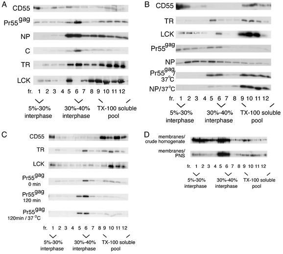
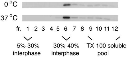
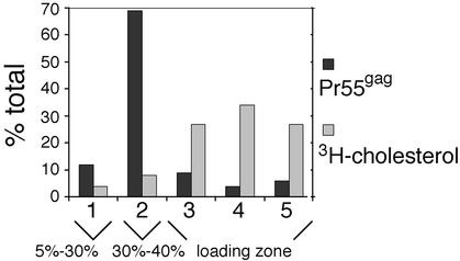
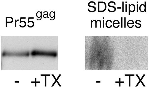
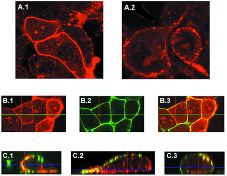
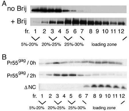
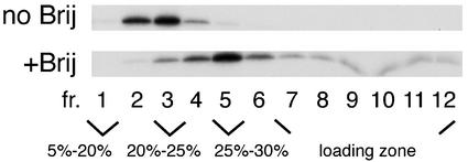
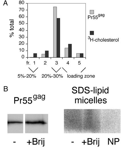
The assembly and budding of human immunodeficiency virus type 1 (HIV-1) at the plasma membrane are directed by the viral core protein Pr55(gag). We have analyzed whether Pr55(gag) has intrinsic affinity for sphingolipid- and cholesterol-enriched raft microdomains at the plasma membrane. Pr55(gag) has previously been reported to associate with Triton X-100-resistant rafts, since both intracellular membranes and virus-like Pr55(gag) particles (VLPs) yield buoyant Pr55(gag) complexes upon Triton X-100 extraction at cold temperatures, a phenotype that is usually considered to indicate association of a protein with rafts. However, we show here that the buoyant density of Triton X-100-treated Pr55(gag) complexes cannot be taken as a proof for raft association of Pr55(gag), since lipid analyses of Triton X-100-treated VLPs demonstrated that the detergent readily solubilizes the bulk of membrane lipids from Pr55(gag). However, Pr55(gag) might nevertheless be a raft-associated protein, since confocal fluorescence microscopy indicated that coalescence of GM1-positive rafts at the cell surface led to copatching of membrane-bound Pr55(gag). Furthermore, extraction of intracellular membranes or VLPs with Brij98 yielded buoyant Pr55(gag) complexes of low density. Lipid analyses of Brij98-treated VLPs suggested that a large fraction of the envelope cholesterol and phospholipids was resistant to Brij98. Collectively, these results suggest that Pr55(gag) localizes to membrane microdomains that are largely resistant to Brij98 but sensitive to Triton X-100, and these membrane domains provide the platform for assembly and budding of Pr55(gag) VLPs.


Figures

FIG. 1.
FIG. 1.
Cold Triton X-100 extraction of cells yields variable complexes for Pr55gag, depending on which cellular fraction is used as a starting material for the detergent treatment. (A) Analyses of crude cell lysates. Pr55gag, CD55, influenza virus NP, the nucleocapsid protein C of SFV, and the TR were expressed in Jurkat cells from recombinant SFV genomes. Cells were extracted with 1% Triton X-100 (TX-100) at 0°C, and lysates were subjected to flotation on iodixanol step gradients. Aliquots of gradient fractions were analyzed by SDS-PAGE. The Pr55gag, NP, and C proteins were analyzed after a 15-min pulse with [35S]methionine and a 60-min chase, and TR was analyzed after a 60-min pulse and 120-min chase, whereas CD55 and the endogenous Lck were detected by Western blotting. (B) Analyses of the total membrane fraction from Jurkat crude cell homogenates. Pr55gag was analyzed after a 15-min pulse with [35S]methionine and a 60-min chase, NP was analyzed after a 60-min pulse, and TR was analyzed after a 60-min pulse and 30-min chase. Total CD55 and Lck were visualized by Western blotting. (C) Analyses of the total membrane fraction from the PNS of Jurkat cells. Pr55gag was analyzed after a 15-min pulse with [35S]methionine and a 0- or 120-min chase, and TR was analyzed after a 90-min pulse. Total CD55 and Lck were visualized by Western blotting. (D) Analyses of membrane-bound Pr55gag from transfected 293T cells. Total Pr55gag in gradient fractions was detected by Western blotting.
FIG. 2.
FIG. 2.
Triton X-100 extraction of VLPs at 0°C or 37°C yields buoyant Pr55gag complexes that float to the 30 to 40% iodixanol interphase. VLPs were collected from SFV-C/HIVgag-infected Jurkat cells which had been metabolically labeled with [35S]methionine for 180 min. VLPs were extracted with 1% Triton X-100 (TX-100) and fractionated on iodixanol step gradients.
FIG. 3.
FIG. 3.
Triton X-100 extraction efficiently strips envelope cholesterol from VLP-Pr55gag. VLPs were collected from SFV-C/HIVgag-infected Jurkat cells that had been metabolically labeled with [35S]methionine or [3H]cholesterol. VLPs were extracted with 1% Triton X-100 at 0°C, and the extracts were fractionated on iodixanol step gradients. Aliquots of gradient fractions were analyzed by SDS-PAGE ([35S]methionine-labeled VLPs) or by scintillation counting ([3H]cholesterol-labeled VLPs). Labeled Pr55gag and [3H]cholesterol in gradient fractions are expressed as percentages of the total (sum of all fractions).
FIG. 4.
FIG. 4.
Triton X-100 extracts the bulk of envelope phospholipids from the VLP-Pr55gag. VLPs were collected from SFV-C/HIVgag-infected Jurkat cells that had been metabolically labeled with [35S]methionine and [32P]orthophosphate. VLPs were extracted with 1% Triton X-100 at 0°C, and the lysate was fractionated on an iodixanol step gradient as shown in Fig. 1. Buoyant Pr55gag complexes from the 30 to 40% iodixanol interphase were concentrated by ultracentrifugation. The resulting pellet (+TX), as well as an intact, non-Triton X-100-extracted VLP sample (−), were solubilized with hot SDS and analyzed by SDS-PAGE. Equivalent amounts of samples were loaded on 10% (Pr55gag) and 20% (SDS-lipid micelles) polyacrylamide gels. On 20% polyacrylamide gels, mixed SDS-lipid micelles separate from other 32P-labeled material. Comparison of the 10 and 20% polyacrylamide gels demonstrates that although the intact VLP and the Triton X-100-treated samples contained similar amounts of Pr55gag complexes, the two samples significantly differed in their phospholipid contents. The radioactive signal in the mixed SDS-lipid micelles originates from both glycerophospholipids and sphingomyelin.
FIG. 5.
FIG. 5.
Confocal fluorescence microscopy of transfected 293T cells. (A) Different anti-Pr55gag antibodies give different staining patterns for Pr55gag at the cell surface. (A.1) MAb EF-7. (A.2) MAb 38:9. (B) Both Pr55gag and GM1 exhibit smooth staining patterns on transfected 293T cells if GM1-containing rafts have not coalesced. Live cells were first incubated with FITC-conjugated cholera toxin B subunit at a cold temperature to detect GM1, and the cells were subsequently fixed, permeabilized, and incubated with MAb EF-7 and TRITC-conjugated antimouse IgG antibodies to detect Pr55gag. (B.1) Pr55gag. (B.2) GM1. (B.3) Pr55gag and GM1 images superimposed. (C) Coalescence of GM1-positive rafts at the cell surface transforms the smooth staining pattern of Pr55gag into a patchy one. Live cells were first incubated with FITC-conjugated cholera toxin B subunit at a cold temperature, and the GM1-positive rafts were subsequently patched by brief incubation at 37°C in the presence of anti-cholera toxin antibodies. Cells were then fixed, permeabilized, and immunostained with MAb EF-7 and TRITC-conjugated antimouse IgG antibodies to detect Pr55gag. Three representative examples of patched cells (C.1 to C.3) are shown with Pr55gag (red) and GM1 (green) images superimposed. Yellow indicates extensive colocalization of Pr55gag and GM1. A single optical section is shown in panels A and B, whereas all optical sections (the Z-stack) were superimposed in panel C and tilted with the orthogonal function to show the view through the whole cell.
FIG. 6.
FIG. 6.
Intracellular membrane-bound Pr55gag is largely resistant to Brij98. (A) Analyses of transfected 293T cells. The total membrane fraction from PNS was extracted with Brij98 for 5 min at 37°C or left untreated, and the samples were subjected to flotation on iodixanol step gradients. Total Pr55gag in gradient fractions was visualized by Western blotting. (B) Brij98 resistance of newly synthesized Pr55gag. The wild type and ΔNC (MA-CA-p2) mutant of Pr55gag were expressed in Jurkat cells from recombinant SFV genomes. Cells were metabolically labeled with [35S]methionine for 15 min and chased for 0 min and 2 h (Pr55gag) or for 2 h (ΔNC). The total membrane fraction from PNS was extracted with Brij98 as described above, and the extracts were fractionated on iodixanol step gradients. Aliquots of gradient fractions were analyzed by SDS-PAGE.
FIG. 7.
FIG. 7.
Brij98-treated VLPs retain low buoyant density. VLPs were collected from SFV-C/HIVgag-infected Jurkat cells that had been metabolically labeled with [35S]methionine for 180 min. The VLPs were extracted with Brij98 and analyzed as described in the legend to Fig. 6. No Brij indicates intact, nonextracted VLPs.
FIG. 8.
FIG. 8.
Large fraction of envelope phospholipids and cholesterol remains attached to Brij98-treated VLP-Pr55gag. (A) Brij98 extraction of [35S]methionine- or [3H]cholesterol-labeled VLPs produced from SFV-C/HIVgag-infected Jurkat cells. The VLPs were extracted with Brij98 for 5 min at 37°C, and the samples were processed as described in the legend to Fig. 3, with the exception that the iodixanol step gradient used was slightly different. (B) Brij98 extraction of [35S]methionine- and [32P]orthophosphate-labeled VLPs produced from SFV-C/HIVgag-infected Jurkat cells. The VLPs were extracted with Brij98 for 5 min at 37°C, and the extracts were fractionated on iodixanol step gradients as described in the legend to Fig. 6. The buoyant Pr55gag complexes from fractions 3 to 5 were pooled and concentrated by pelletation. The pellets, as well as intact, nonextracted VLP samples (−) were analyzed as described in the legend to Fig. 4. NP is a Brij98-treated control sample that was collected from [32P]orthophosphate-labeled Jurkat cells infected with recombinant SFV encoding influenza virus NP. The NP control demonstrates that the majority of phospholipid signal in the Brij98-treated VLP sample originates from VLPs, not from contaminating microvesicles.

References

    1. Aliperti, G., and M. J. Schlesinger. 1978. Evidence for an autoprotease activity of Sindbis virus capsid protein. Virology 90:366-369. - PubMed
    1. Aloia, R. C., F. C. Jensen, C. C. Curtain, P. W. Mobley, and L. M. Gordon. 1988. Lipid composition and fluidity of the human immunodeficiency virus. Proc. Natl. Acad. Sci. USA 85:900-904. - PMC - PubMed
    1. Aloia, R. C., H. Tian, and F. C. Jensen. 1993. Lipid composition and fluidity of the human immunodeficiency virus envelope and host cell plasma membranes. Proc. Natl. Acad. Sci. USA 90:5181-5185. - PMC - PubMed
    1. Benting, J. H., A. G. Rietveld, and K. Simons. 1999. N-glycans mediate the apical sorting of a GPI-anchored, raft-associated protein in Madin-Darby kidney cells. J. Cell Biol. 146:313-320. - PMC - PubMed
    1. Brown, D. A. 1998. Structure and origin of ordered lipid domains in biological membranes. J. Membr. Biol. 164:103-114. - PubMed

Publication types

LinkOut - more resources